

Orthotopic pituitary tumors generated by stereotaxic GC cell injection in immunocompetent rats
- PMID: 40618150
- PMCID: PMC12229040
- DOI: 10.1186/s40478-025-02052-6
Orthotopic pituitary tumors generated by stereotaxic GC cell injection in immunocompetent rats
Abstract
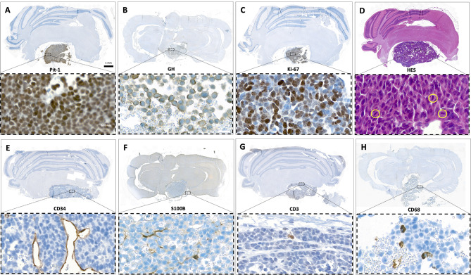
Innovative treatment strategies for pituitary tumors are necessary to limit the disease burden and to improve survival in cases of carcinomas. The paucity and inaccuracy of available preclinical models substantially hamper pituitary research and drug discovery. Hence, we describe a novel method to generate orthotopic pituitary tumors via stereotaxic injection of somatotroph GC cells into the pituitaries of immunocompetent Wistar Furth rats. Tumor growth was monitored by repeated 7 Tesla magnetic resonance imaging. The procedure consistently led to rapidly expanding intra- and suprasellar growth hormone-secreting tumors within their native anatomical environment. The generated tumors faithfully reproduced the microarchitecture of human somatotroph pituitary adenomas, including the immune infiltrates and other typical components of their microenvironment, which is a prerequisite for testing immunomodulating agents. This orthotopic model of proliferative pituitary tumors developed in immunocompetent hosts therefore unlocks new opportunities for preclinical studies.
Keywords: Immunocompetent rat; Pituitary adenoma; Pituitary research; Preclinical model; Tumor microenvironment.
© 2025. The Author(s).
Conflict of interest statement
Declarations. Ethics approval and consent to participate: All experiments were conducted in compliance with the European Union recommendations (2013/63/EU) and were approved by the French Ministry of Higher Education, Research and Innovation (APAFIS #2020022115123683) and the local ethical committee of the Paris-Saclay University. Consent for publication: Not applicable. Competing interests: The authors declare no competing interests. List of abbreviations: none.
Figures


Similar articles
-
Relationship between Pituitary Gland and Stem Cell in the Aspect of Hormone Production and Disease Prevention: A Narrative Review.Endocr Metab Immune Disord Drug Targets. 2025;25(7):509-526. doi: 10.2174/0118715303314551241031093717. Endocr Metab Immune Disord Drug Targets. 2025. PMID: 39812047 Review.
-
Development of orthotopic mouse models for mid-low rectal cancer.Acta Pharmacol Sin. 2025 Jun;46(6):1772-1781. doi: 10.1038/s41401-025-01489-8. Epub 2025 Feb 12. Acta Pharmacol Sin. 2025. PMID: 39939805
-
A multiparametric perspective on C6 and F98 cell lines in orthotopic rat models for glioblastoma research.Sci Rep. 2025 Jul 2;15(1):22547. doi: 10.1038/s41598-025-06684-5. Sci Rep. 2025. PMID: 40596200 Free PMC article.
-
A contemporary, multiinstitutional analysis of transcription factor lineage in pituitary adenomas: comparative study of neuroimaging, histopathology, and clinical outcomes.J Neurosurg. 2025 Mar 14;143(1):146-154. doi: 10.3171/2024.10.JNS24853. Print 2025 Jul 1. J Neurosurg. 2025. PMID: 40085941
-
Metabolic In Vivo Visualization of Pituitary Adenomas: a Systematic Review of Imaging Modalities.World Neurosurg. 2017 Aug;104:489-498. doi: 10.1016/j.wneu.2017.04.128. Epub 2017 Apr 28. World Neurosurg. 2017. PMID: 28461279 Free PMC article.
References
-
- Gittleman H, Ostrom QT, Farah PD, Ondracek A, Chen Y, Wolinsky Y et al (2014) Descriptive epidemiology of pituitary tumors in the united states, 2004–2009. J Neurosurg 121:527–535 - PubMed
-
- Melmed S, Pituitary-Tumor Endocrinopathies (2020) N Engl J Med 382:937–950 - PubMed
-
- Gengenbacher N, Singhal M, Augustin HG (2017) Preclinical mouse solid tumour models: status quo, challenges and perspectives. Nat Rev Cancer 17:751–765 - PubMed
-
- Heaney AP, Fernando M, Yong WH, Melmed S (2002) Functional PPAR-γ receptor is a novel therapeutic target for ACTH-secreting pituitary adenomas. Nat Med 8:1281–1287 - PubMed
MeSH terms
LinkOut - more resources
Full Text Sources
Medical
Miscellaneous
