. 2025 Jul 4:1-14.
doi: 10.1159/000547257. Online ahead of print.

Mesenchymal Stromal Cells from Dental Tissues Demonstrate Neuronal Potential Pre- and Post-Induction

Affiliations

Mesenchymal Stromal Cells from Dental Tissues Demonstrate Neuronal Potential Pre- and Post-Induction

Agner Henrique Dorigo Hochuli et al. Cells Tissues Organs. .

Abstract

Introduction: Mesenchymal stromal cells (MSCs) play a crucial role in tissue repair and exhibit anti-inflammatory properties, making them promising for regenerative medicine. Dental tissue-derived MSCs, such as stromal cells from human exfoliated deciduous teeth (SHEDs) and dental pulp stromal cells (DPSCs), express neural markers and hold the potential for treating neurodegenerative diseases. Nonetheless, their relative ability to differentiate into neurons is still unclear. Given their shared embryonic origin, we hypothesised that SHEDs and DPSCs possess similar potential for neuronal differentiation along with intrinsic expression of neuronal markers. The objective of this study was to compare their differentiation abilities under standardised conditions, evaluate neuronal markers pro- and post-neuronal induction, and compare the neuronal differentiation potential of SHEDs and DPSCs.

Methods: SHEDs (n = 3) and DPSCs (n = 3) were collected with ethical approval, cultured, and characterised according to established MSC criteria. Clonogenicity, proliferation, senescence, and trilineage differentiation were assessed. Neuronal differentiation was induced for 21 days and evaluated using flow cytometry (SRY-Box Transcription Factor 1 [SOX1], SRY-Box Transcription Factor 2 [SOX2], glial fibrillary acidic protein [GFAP], doublecortin, nestin, CD56, CD146), immunofluorescence for βIII-tubulin, reverse transcription polymerase chain reaction for tubulin 3 (TUB3), and microtubule-associated protein 2 (MAP2).

Results: Both SHEDs and DPSCs exhibited MSC characteristics. SHEDs showed higher clonogenicity. Early neuronal markers (e.g., SOX1, nestin, GFAP, βIII-tubulin) were detected pre- and post-induction in both cell types without significant intergroup differences. No significant expression of TUB3 and MAP2 was observed.

Conclusion: SHEDs and DPSCs show comparable neuronal marker expression profiles, suggesting similar early neuronal differentiation potential. These findings support using undifferentiated SHEDs and DPSCs in neuroregenerative strategies, offering cost-effective and safer alternatives to pre-differentiated cells.

Keywords: Dental tissues; Mesenchymal stem cells; Neuronal differentiation; Regenerative medicine; Stem cell differentiation.


Conflict of interest statement

The authors declared no potential conflicts of interest with respect to the research, authorship, and/or publication of this article.

Figures
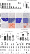
Fig. 1.
Fig. 1.
Characterisation of MSCs derived from exfoliated deciduous teeth (SHED) and dental pulp MSC derived from permanent teeth (DPSC). a Flow cytometry reveals mean and percentage expression of MSC markers in SHED and DPSC. b STRO-1 expression in SHED and DPSC. c In vitro differentiation – representative image. Osteogenic lineage: control; differentiation; red calcium crystals (stained with Alizarin Red S). Adipogenic lineage: control; differentiation; red lipid droplets (stained with oil red O). Chondrogenic lineage: control; differentiation; cuboidal cells; and gaps around young chondrocytes (stained with toluidine blue). Magnification, ×400. Scale bar, 50 μm. d Adipogenic differentiation quantified by microplate reader at 550 nm OD. e Osteogenic differentiation quantified by microplate reader at 450 nm OD. f Representative karyogram of SHEDs after cell expansion with a normal karyotype: 46,XY. g Representative karyogram of DPSCs after cell expansion presenting a normal karyotype: 46,XX. Data presented as mean ± SD. SHEDa, exfoliated primary tooth pulp stem cells induced to adipogenic differentiation; DPSCa, permanent tooth pulp stem cells induced to adipogenic differentiation; SHEDo, exfoliated primary tooth pulp stem cells induced to ostegenic differentiation; DPSCo, permanent tooth pulp stem cells induced to ostegenic differentiation.
Fig. 2.
Fig. 2.
Clonogenic, senescence, and proliferation potential of MSCs derived from exfoliated deciduous teeth (SHED) and dental pulp MSC derived from permanent teeth (DPSC). a Representative crystal violet-stained images of SHED and DPSC fibroblast colony-forming units (CFU-F) (magnification, ×40). b Graph depicting CFU-F for SHED and DPSC, along with colony-forming efficiency after 10 days of culture. c Comparative analysis of SHED and DPSC in cell proliferation assay, measuring EdU expression by flow cytometry at passages three (P3), five (P5), and seven (P7). d Comparative analysis of SHED and DPSC in cell senescence assay, assessing β-galactosidase expression by flow cytometry at passages three (P3), six (P6), nine (P9), and twelve (P12). Data are presented as mean ± standard deviation. The p value was obtained from paired Student’s t test.
Fig. 3.
Fig. 3.
Expression of neuronal markers in MSCs derived from exfoliated deciduous teeth (SHED) and dental pulp MSC derived from permanent teeth (DPSC). a Immunofluorescence representative images of SHED pre- and post-neuronal induction labelled with anti-βIII-tubulin (green marker). The nucleus is blue (DAPI). ×400, magnification. Scale bar, 50 μm. b Immunofluorescence representative images of DPSC pre- and post-neuronal induction labelled with anti-βIII-tubulin (green marker). The nucleus is blue (DAPI). ×400, magnification. Scale bar, 50 μm. c Quantification of the fluorescence intensity of βIII-tubulin in SHED and DPSC pre- and post-neuronal induction. d Expression of tubulin 3 (TUB3) and microtubule-associated protein 2 (MAP2) in SHED and DPSC pre- and post-neuronal induction and neural stem cells (NSC) (used as positive control) by reverse transcription polymerase chain reaction (RT-PCR) in 2% agarose gel. The glyceraldehyde-3-phosphate dehydrogenase (GAPDH) was used as a constitutive housekeeping gene to normalise changes in specific gene expression. The p value was obtained from the ANOVA test.

References

    1. Caplan AI. Why are MSCs therapeutic? New Data: New Insight J Pathol. 2009;217(2):318–24. - PMC - PubMed
    1. Dominici M, Le Blanc K, Mueller I, Slaper-Cortenbach I, Marini FC, Krause DS, et al. Minimal criteria for defining multipotent mesenchymal stromal cells. The International Society for cellular therapy position statement. Cytotherapy. 2006;8(4):315–7. - PubMed
    1. Shi S, Bartold P, Miura M, Seo B, Robey P, Gronthos S. The efficacy of mesenchymal stem cells to regenerate and repair dental structures. Orthod Craniofac Res. 2005;8(3):191–9. - PubMed
    1. Gronthos S, Brahim J, Li W, Fisher LW, Cherman N, Boyde A, et al. Stem cell properties of human dental pulp stem cells. J Dent Res. 2002;81(8):531–5. - PubMed
    1. Victor AK, Reiter LT. Dental pulp stem cells for the study of neurogenetic disorders. Hum Mol Genet. 2017;26(R2):R166–71. - PMC - PubMed