

HOXD13 knockdown attenuates apoptosis in prostate cancer via the upregulation of MDM2/p53 signaling
- PMID: 40619412
- PMCID: PMC12232685
- DOI: 10.1186/s13008-025-00161-1
HOXD13 knockdown attenuates apoptosis in prostate cancer via the upregulation of MDM2/p53 signaling
Abstract
Background: Prostate cancer (PCa) is a common leading cause of death worldwide and is recognized as the second most frequently diagnosed cancer in the male population, making it a significant focus of long-term oncological research. The HOXD13 gene, belonging to the HOX gene family, has been associated with various malignancies, including breast and colon cancer, and is associated with prognostic outcomes. However, its specific role in prostate cancer remains to be elucidated.
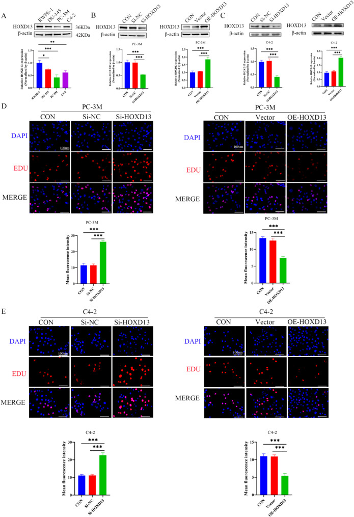
Methods: The correlation of HOXD13 expression in PCa was analyzed by UALCAN database. qRT-PCR and western blot assays were employed to assess the expression levels of HOXD13 in several prostate cancer cell lines. Construct siRNA and overexpression plasmids for HOXD13 and transfect them into cells. Western blot was used to assess the knockdown efficiency of HOXD13 in the cells. The influence of HOXD13 expression on cell proliferation in PC-3 M and C4-2 cells was assessed by EDU staining. The role of HOXD13 in cell migration and invasion of prostate cancer cells was detected by wound healing and transwell assay. Western blot analysis was performed to examine apoptosis-related proteins (C-caspase3, Bax, and Bcl-2) in PC-3 M cells. The co-immunoprecipitation (Co-IP) assay evaluates the interaction between MDM2 and p53.
Results: An examination of the data obtained from the UALCAN database reveals that HOXD13 was significantly reduced in prostate cancer tissues and correlated with patient survival. In prostate cancer cell lines, notably PC-3M, HOXD13 expression was markedly reduced. Knockdown of HOXD13 promotes PC-3 M and C4-2 cells proliferation, migration, and invasion. However, the elevated expression of HOXD13 hampers PC-3 M and C4-2 cells proliferation, migration, and invasion. Furthermore, knockdown of HOXD13 in PC-3 M cells resulted in decreased levels of apoptosis markers, including C-caspase-3 and Bax. Co-immunoprecipitation assays demonstrated that HOXD13 depletion enhanced the association between MDM2 and p53.
Conclusion: HOXD13 was a critical regulator in the pathogenesis of prostate cancer, modulating the MDM2/p53 signaling pathway to promote apoptosis.
Keywords: HOXD13; Apoptosis; MDM2; Prostate cancer; p53.
© 2025. The Author(s).
Conflict of interest statement
Declarations. Ethics approval and consent to participate: Not applicable. Competing interests: The authors declare no competing interests.
Figures


Similar articles
-
Caveolin-1 inhibits the proliferation and invasion of lung adenocarcinoma via EGFR degradation.Sci Rep. 2025 Jul 1;15(1):21654. doi: 10.1038/s41598-025-05259-8. Sci Rep. 2025. PMID: 40594106 Free PMC article.
-
Prescription of Controlled Substances: Benefits and Risks.2025 Jul 6. In: StatPearls [Internet]. Treasure Island (FL): StatPearls Publishing; 2025 Jan–. 2025 Jul 6. In: StatPearls [Internet]. Treasure Island (FL): StatPearls Publishing; 2025 Jan–. PMID: 30726003 Free Books & Documents.
-
SHCBP1 promotes tumor cell proliferation, migration, and invasion, and is associated with poor prostate cancer prognosis.J Cancer Res Clin Oncol. 2020 Aug;146(8):1953-1969. doi: 10.1007/s00432-020-03247-1. Epub 2020 May 23. J Cancer Res Clin Oncol. 2020. PMID: 32447485 Free PMC article.
-
[Volume and health outcomes: evidence from systematic reviews and from evaluation of Italian hospital data].Epidemiol Prev. 2013 Mar-Jun;37(2-3 Suppl 2):1-100. Epidemiol Prev. 2013. PMID: 23851286 Italian.
-
Cost-effectiveness of using prognostic information to select women with breast cancer for adjuvant systemic therapy.Health Technol Assess. 2006 Sep;10(34):iii-iv, ix-xi, 1-204. doi: 10.3310/hta10340. Health Technol Assess. 2006. PMID: 16959170
References
-
- Salsi V, Zappavigna V. Hoxd13 and Hoxa13 directly control the expression of the EphA7 Ephrin tyrosine kinase receptor in developing limbs. J Biol Chem. 2006;281(4):1992–9. - PubMed
-
- Cantile M, Franco R, Tschan A, et al. HOX D13 expression across 79 tumor tissue types. Int J Cancer. 2009;125(7):1532–41. - PubMed
-
- Apiou F, Flagiello D, Cillo C, Malfoy B, Poupon MF, Dutrillaux B. Fine mapping of human HOX gene clusters. Cytogenet Cell Genet. 1996;73(1–2):114–5. - PubMed
-
- Scott MP. Vertebrate homeobox gene nomenclature. Cell. 1992;71(4):551–3. - PubMed
Grants and funding
- No. 20240043/Medical Science Research Project of Hebei Provincial Health Commission
- No. 20240043/Medical Science Research Project of Hebei Provincial Health Commission
- No. 20240043/Medical Science Research Project of Hebei Provincial Health Commission
- No. 20240043/Medical Science Research Project of Hebei Provincial Health Commission
- No. 20240043/Medical Science Research Project of Hebei Provincial Health Commission
LinkOut - more resources
Full Text Sources
Research Materials
Miscellaneous
