Review
. 2025 Jul 6;13(1):93.
doi: 10.1186/s40364-025-00805-y.

Proteomics in pancreatic cancer

Affiliations
Review

Proteomics in pancreatic cancer

Fei Cai et al. Biomark Res. .

Abstract

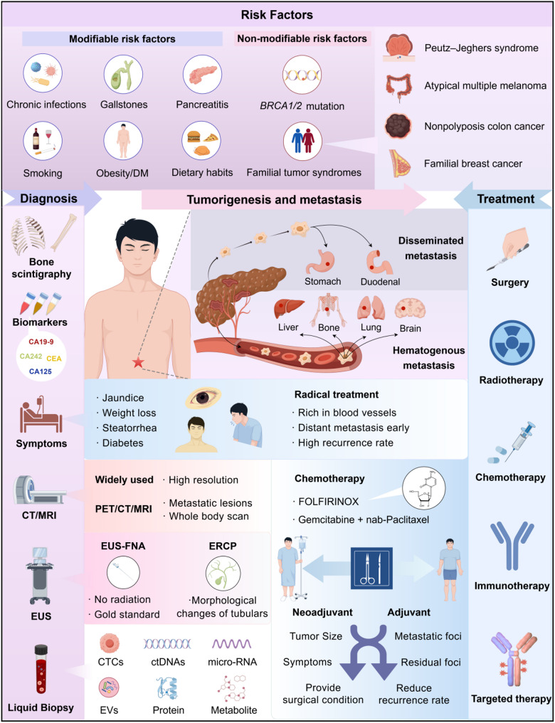
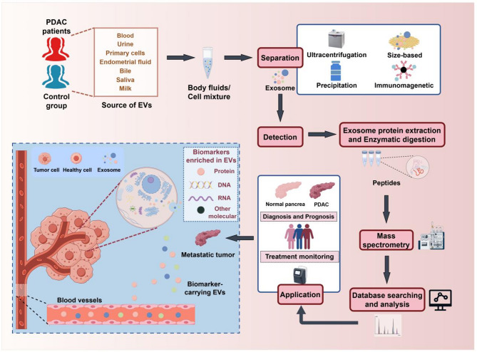
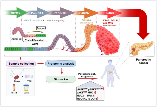
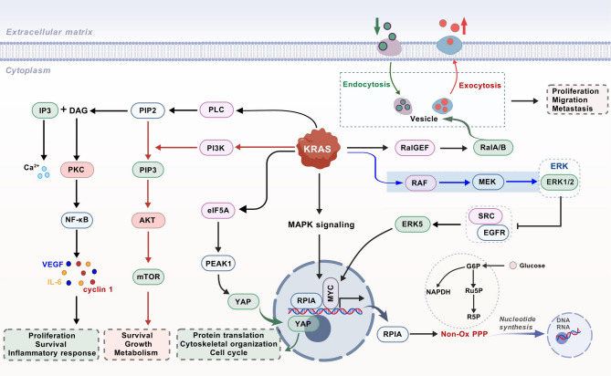
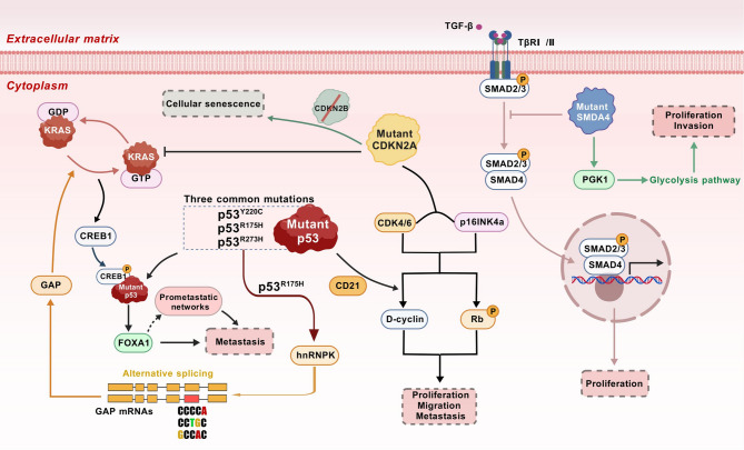
Pancreatic cancer (PC), one of the most aggressive malignancies, is characterized by a dismal prognosis owing to its low early detection rates, rapid progression, frequent postoperative complications, and limited efficacy of conventional oncological therapies. The fact that most patients are diagnosed at advanced stages underscores the critical importance of early detection for the formulation of effective treatment strategies. Despite substantial research efforts, the medical community still lacks consistent and dependable biomarkers for the diagnosis, classification, and prognosis of PC, highlighting the urgent need for innovative and more efficient approaches to identify pancreatic abnormalities at early stages. For decades, mass spectrometry (MS)-based proteomics has been extensively applied in disease diagnostics, mechanistic investigations, and screening of potential drug targets. This review systematically synthesizes recent advancements in clinical proteomic techniques and applications, highlighting significant biomarker discoveries and signal transduction networks associated with PC. By integrating these findings, we provide novel insights into the molecular mechanisms underlying PC development and progression, which may facilitate the identification of new diagnostic biomarkers and therapeutic targets for this disease.

Keywords: Biomarkers; Mass spectrometry; Pancreatic cancer; Pancreatic ductal adenocarcinoma; Proteomics.


Conflict of interest statement

Declarations. Ethics approval and consent to participate: Not applicable. Consent for publication: Not applicable. Competing interests: The authors declare no competing interests.

Figures

Fig. 1
Fig. 1
Comprehensive overview of PC diagnosis and treatment. In recent years, significant advancements have been made in diagnostic technologies for PC. In addition to traditional imaging techniques and pathological diagnosis methods, liquid biopsies and artificial intelligence methods are gradually being integrated into clinical practice. Treatment options for PC vary, with recent clinical studies highlighting the importance of individualized and precision treatments. Long-term follow-up management for PC patients is also essential, as it can influence therapeutic outcomes and enhance patients’ quality of life. Abbreviations: CT, computed tomography; MRI, magnetic resonance imaging; EUS, endoscopic ultrasonography; ERCP, endoscopic retrograde cholangiopancreatography; CTCs, circulating tumor cells; ctDNA, circulating tumor DNA; EVs, extracellular vesicles. The figure was created by Figdraw (www.figdraw.com)
Fig. 2
Fig. 2
Significant milestones and breakthroughs in proteomics. Abbreviations: 2D-PAGE, two-dimensional polyacrylamide gel electrophoresis; ESI, electrospray ionization; MALDI-TOF-MS, matrix-assisted laser desorption/ionization time‒of‒flight mass spectrometry; HPA, human protein atlas; HUPO, human proteome organization; LC‒MS/MS, liquid chromatography‒tandem mass spectrometry; HPP, human proteome project; HCC, hepatocellular carcinoma
Fig. 3
Fig. 3
Schematic illustration of the PC proteomic workflow. Samples are first collected from both in vivo and in vitro sources. The samples subsequently undergo lysis to extract proteins, which are then enzymatically digested into peptide mixtures. These peptides are analyzed via LC‒MS/MS in either DDA or DIA acquisition mode. The acquired data are processed and visualized through database searching and bioinformatics analysis. Statistically significant proteins are then selected through rigorous statistical analysis. Finally, these candidate proteins are further validated in clinical populations via integration with clinical data for potential clinical translation. Abbreviations: FFPE, formalin-fixed paraffin-embedded; LC‒MS/MS, liquid chromatography‒tandem mass spectrometry; DDA, data-dependent acquisition; DIA, data-independent acquisition. The figure was created with BioGDP.com (https://BioGDP.com)
Fig. 4
Fig. 4
Mass spectrometry-based workflow for extracellular vesicle proteomics. PDAC-derived sEVs that circulate in the blood can be enriched by techniques such as ultracentrifugation. Molecular components, including proteins, can be analyzed to generate unique biomarkers for PDAC diagnosis/prognosis. Abbreviations: PDAC, pancreatic ductal adenocarcinoma; EVs, extracellular vesicles. The figure was created by Figdraw (www.figdraw.com)
Fig. 5
Fig. 5
Underlying mechanisms of several potential PC therapeutics via proteomic approaches. On the one hand, for compounds with established antitumor activity but undefined mechanisms in PC, deep-coverage proteomics enables systematic screening of differential protein signatures. This approach facilitates the precise identification of molecular targets and associated mechanistic pathways. On the other hand, for candidate compounds with therapeutic potential in PC with unknown targets, proteomic profiling provides a powerful platform to delineate pharmacodynamic biomarkers and elucidate the underlying mode of action. Some potential drugs and their related mechanisms of action are currently being explored in the context of PDAC and are shown in the figure. “↑” indicates activation, stimulation or promotion, whereas “⊥” indicates inhibition, suppression or decrease. The figure was created with BioGDP.com (https://BioGDP.com)
Fig. 6
Fig. 6
Mechanisms for initiating PanINs and their progression to PDAC. Oncogenic KRAS induces neoplastic transformation of pancreatic acinar cells through acinar-to-ductal metaplasia (ADM), an actin-based morphogenetic process, and drives PDAC. During the progression from low-grade to high-grade precursor lesions, inactivation of the tumor suppressor gene CDKN2A is observed. As these precursor lesions progress to PDAC, concomitant loss or inactivation mutations in the critical tumor suppressor genes BRCA2 or TP53 occur. Longitudinal collection of stage-stratified patient samples (e.g., tissue or liquid biopsies) during this progression, coupled with proteomic approaches, may enable systematic screening of biomarkers possessing dual early diagnostic and prognostic potential, thereby enhancing prognostic stratification capabilities in PDAC management. FFPE, formalin-fixed paraffin-embedded. The figure was created with BioGDP.com (https://BioGDP.com)
Fig. 7
Fig. 7
Some KRAS signaling pathways mediating PC mentioned in this review. KRAS is activated through the binding of ligands and receptors and transmits signals to multiple downstream signaling pathways. On the one hand, it affects endocytosis, exocytosis and the distribution of receptors on the cell membrane. On the other hand, it regulates the expression of various genes in the nucleus, playing a role in multiple physiological processes in tumor cells. “↑” indicates activation, stimulation or promotion, whereas “⊥” indicates inhibition, suppression or decrease. Abbreviations: PLC, phospholipase C; PI3K, phosphoinositide 3-kinase; PIP3, phosphatidylinositol 3,4,5-trisphosphate; NF-κB, nuclear factor kappa-B; mTOR, mammalian target of rapamycin; eIF5A, eukaryotic translation initiation factor 5 A; PEAK1, pseudopodium enriched atypical kinase 1; YAP, myelocytomatosis oncogene; MEK, mitogen-activated protein kinase/extracellular signal-regulated kinase; RAF, Proto-Oncogene Serine/Threonine-Protein Kinase; ERK, extracellular regulated kinase; RPIA, ribose 5-phosphate isomerase A; Non-Ox PPP, the non-oxidative oxidative branch of the pentose phosphate pathway; Ru5P, ribulose 5-phosphate; G6P, glucose-6-phosphatase G-6-pase. The figure was created with BioGDP.com (https://BioGDP.com)
Fig. 8
Fig. 8
Some p53, CDKN2A, and SMAD4 signaling pathways mediating PC mentioned in this review. P53 is a crucial tumor suppressor gene. The figure illustrates its three most common mutation forms. It influences the metabolism, metastasis, and proliferation of tumor cells through downstream signaling molecules. It interacts with the KRAS signaling pathway through hnRNPK and CREB and with CDKN2A via CD21. Mutated CDKN2A generates the p16 biomarker, which affects genomic stability and cell proliferation through cyclins and Rb. SMAD4 affects cell proliferation by regulating the expression of genes related to TGF-β in the nucleus. In the cytoplasm, it impacts the cell glycolysis process via PGK1 and influences cell proliferation and tumor growth. “↑” indicates activation, stimulation or promotion, whereas “⊥” indicates inhibition, suppression or decrease. Abbreviations: TGF-β, transforming growth factor β; SMAD, small mothers against decapentaplegic homolog; PGK1, phosphoglycerate kinase 1; CDKN2A, cyclin-dependent kinase inhibitor 2 A; CDK, cyclin-dependent kinase; CREB, cAMP response element binding protein; FOXA1, forkhead box A1; hnRNPK, heterogeneous nuclear ribonucleoprotein K; Rb, retinoblastoma protein. The figure was created with BioGDP.com (https://BioGDP.com)

References

    1. Leiphrakpam PD, Chowdhury S, Zhang M, Bajaj V, Dhir M, Are C. Trends in the global incidence of pancreatic Cancer and a brief review of its histologic and molecular subtypes. Journal Gastrointest cancer. 2025;24(1):71. 10.1007/s12029-025-01183-2. - PubMed
    1. Siegel RL, Kratzer TB, Giaquinto AN, Sung H, Jemal A. Cancer statistics, 2025. CA Cancer J Clin. 2025;75(1):10–45. 10.3322/caac.21871. - PMC - PubMed
    1. Kleeff J, Korc M, Apte M, et al. Pancreatic cancer. Nat Rev Dis Primers. 2016;21:2:16022. 10.1038/nrdp.2016.22. - PubMed
    1. Nevala-Plagemann C, Hidalgo M, Garrido-Laguna I. From state-of-the-art treatments to novel therapies for advanced-stage pancreatic cancer. Nat Rev Clin Oncol. 2020;17(2):108–23. 10.1038/s41571-019-0281-6. - PubMed
    1. Ho WJ, Jaffee EM, Zheng L. The tumour microenvironment in pancreatic cancer - clinical challenges and opportunities. Nat Rev Clin Oncol. 2020;17(9):527–40. 10.1038/s41571-020-0363-5. - PMC - PubMed

LinkOut - more resources