. 2025 Jul;14(7):e70113.
doi: 10.1002/jev2.70113.

Defining the Parameters for Sorting of RNA Cargo Into Extracellular Vesicles

Affiliations

Defining the Parameters for Sorting of RNA Cargo Into Extracellular Vesicles

Ahmed Abdelgawad et al. J Extracell Vesicles. 2025 Jul.

Abstract

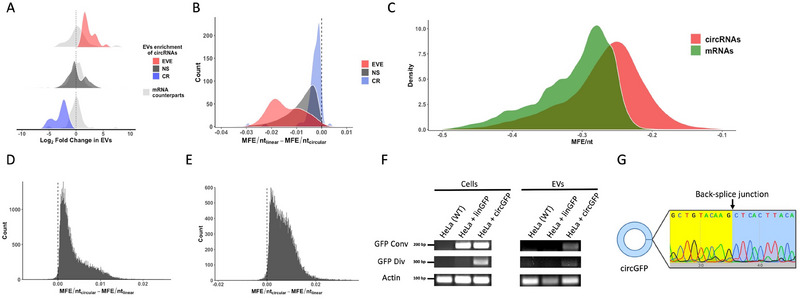
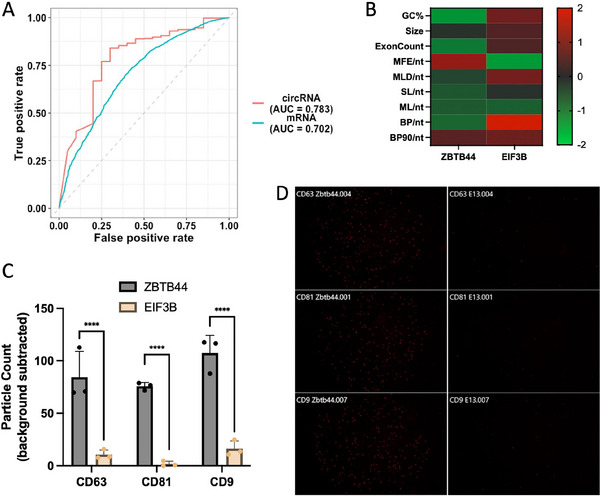
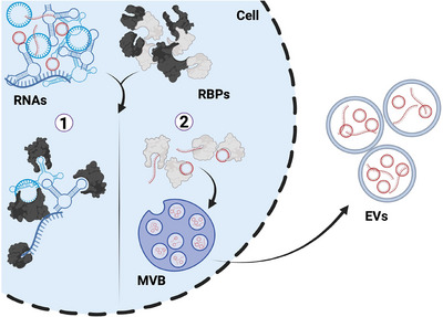
Extracellular vesicles (EVs) are small particles that are released by cells and mediate cell-cell communication by transferring bioactive molecules such as RNA. RNA cargo of EVs, including coding and non-coding RNAs, can change the behaviour of recipient cells, affecting processes including gene expression, proliferation, and Fapoptosis. CircRNAs are stable and resistant to degradation and have been shown to be enriched in EVs. They play key roles in gene regulation and are also emerging as promising biomarkers for disease diagnosis due to their stability and disease-specific expression. Although microRNAs (miRNAs) are the most well studied RNA cargo of EVs, very little is known about the mechanisms of enrichment of circular RNAs (circRNAs) as well as long linear RNAs. Here, we take a comprehensive genome-wide approach to investigate the role of structuredness and shape along with GC%, size, exon count and coding potential, in the sorting and enrichment of circular and long linear RNAs into EVs. We developed a model using these parameters to predict the likelihood of EV packaging of RNA and it was validated by using single molecule RNA imaging of EV bound RNAs. Furthermore, we found that structuredness could explain the relative enrichment of circRNAs over their linear counterparts. These results were validated on existing public databases of circular and linear RNAs in EVs. By identifying and analysing these factors, we aim to better understand the complex mechanisms behind EV-mediated RNA transfer and its impact on cell communication in both health and disease. This mechanistic understanding of RNA enrichment in EVs is crucial for engineering EVs with selective RNA cargo.

Keywords: RNA imaging; SPIRFISH; circRNAs; cis elements; enriched; exosomes; extracellular vesicles; lncRNAs; mRNAs.


Conflict of interest statement

Y.Y. is a named inventor on a patent application (PCT/US2023/020215) for the E3filters used in this study. K.W.W. is or has been an advisory board member of ShiftBio, Exopharm, NeuroDex, NovaDip, and ReNeuron; holds stock options with NeuroDex; and privately consults as Kenneth Witwer Consulting. The authors declare no conflicts of interest. The funders had no role in the design of the study; in the collection, analyses, or interpretation of data; in the writing of the manuscript; or in the decision to publish the results.

Figures

FIGURE 1
FIGURE 1
CircRNAs are less structured compared to their linear counterparts: (A) Average motif count difference between exoRBase circRNAs and the 3’ UTR of their linear counterparts for five RBPs. Motifs of RBPs are shown in brackets. (B) Distribution of size‐normalized structure parameters of circRNAs in exoRBase and their cognate linear isoforms. (C) Change in structure parameters of exoRBase circRNAs after in‐silico linearization. (D) Violin plot of size‐normalized structure parameters of exoRBase circRNAs with high detection frequency compared to those with low frequency. (E) Violin plot of size distribution of exoRBase circRNAs with high detection frequency compared to those with low frequency. Structure parameters are MFE: minimum free energy; MLD: maximum ladder distance; SL: number of stems, ML: number of multiloops of order 3 or higher; BP: Number of paired bases; BP90: 90th percentile of pair distance. p values were calculated using Mann–Whitney's test, NS: no significant difference, *p < 0.05, **p < 0.01, ***p < 0.001.
FIGURE 2
FIGURE 2
CircRNAs are enriched in EVs: (A) Workflow of EVs isolation and RNA‐Seq. (B) Average circular/linear RNA fraction in cell and EVs. (C) Overlap of identified circRNAs with exoRBase circRNAs. (D) Distribution of circRNA exon numbers in cells and EVs. (E) Total number of transcripts detected in either one, two, or all three EV samples from RNA‐Seq for all four classes of RNAs analysed. (F) Ratio of significantly enriched transcripts in EVs compared to cell‐retained transcripts for all four classes of RNAs analysed. (G) circRNAs abundance from RNA‐Seq in EVs and cells. Arrows point to names of circRNAs that were validated by RT‐qPCR. (H) EVs/cell fold Change of circRNAs from three independent samples. CR, cell retained; EVE, EVs enriched; ND, not detected. p values were calculated using Mann–Whitney's test, ***p < 0.001.
FIGURE 3
FIGURE 3
Role of cis elements in enriching circRNAs in EVs: (A) Average motif count difference between cells and EVs circRNAs and the 3’ UTR of their linear counterparts for five RBPs. Motifs of RBPs are shown in brackets. (B) Percentage of EVs enriched (EVE), cell retained (CR), or circRNAs with no significant difference (NS) that contain RBPs motifs. Motifs of RBPs are shown in brackets. (C) Violin plot of size‐normalized structure parameters of EVE and CR circRNAs. (D) Violin plot of size distribution of EVE, NS and CR circRNAs. Structure parameters are MFE: minimum free energy; MLD: maximum ladder distance; SL: number of stems, ML: number of multiloops of order 3 or higher; BP: Number of paired bases; BP90: 90th percentile of pair distance. p values were calculated using Mann–Whitney's test, NS: no significant difference, *p < 0.05, **p < 0.01, ***p < 0.001.
FIGURE 4
FIGURE 4
Role of cis elements in enriching linear RNAs in EVs: (A) mRNAs abundance from RNA‐Seq in EVs and cells. Arrows point to names of mRNAs that were validated by RT‐qPCR. (B) EVs/cell fold change of mRNAs from three independent samples. ND = not detected. (C, D) Similar to A–B but for lncRNAs. (E) Violin plot of size‐normalized structure parameters of EVs enriched (EVE), cell retained (CR), and linear RNAs with no significant difference (NS). (F) Violin plot of size distribution of EVE, NS and CR linear RNAs. (G) Log protein cellular abundance of EVE, NS and CR mRNAs. (H) Gene Ontology (GO) biological process enrichment analysis for EVs enriched mRNAs. p values were calculated using Mann–Whitney's test, ***p < 0.001.
FIGURE 5
FIGURE 5
Circularization enhances RNAs enrichment in EVs: (A) Density plot of log2 fold change in EVs of circRNAs that are EVs enriched (EVE), cell retained (CR), or those with no significant difference (NS) as well as their respective mRNA counterparts. (B) Change in structuredness (MFE) after linearization of EVE, NS or CR circRNAs sequences. (C) Density plot of the structuredness of circRNAs and their mRNA counterparts. (D‐E) Change in structuredness (MFE) after circularization of mRNAs and lncRNAs sequences, respectively. (F) Gel image of WT HeLa cells, or cells expressing linear GFP or circular GFP (circGFP) in cells and EVs using both convergent and divergent primers. (G) Sequence of circGFP covering the back‐splice junction.
FIGURE 6
FIGURE 6
Prediction of RNA enrichment in EVs: (A) Receiver operating characteristic (ROC) curve for EV enrichment prediction models for circRNAs and mRNAs. AUC is shown in brackets. (B) Heatmap of z‐scores of sequence features of ZBTB44 and EIF3B mRNAs. (C) Grouped bar graph comparing the number of CD81+ particles (determined by capture spot) that are detected by interferometry and additionally are determined to be smFISH+. Bars show the mean particle counts across three independent SP‐IRIS microchip experiments; error bars represent standard deviation. Statistical testing was performed by ordinary one‐way ANOVA with Šídák's multiple comparisons test. ****, p ≤ 0.0001. (D) Representative red channel fluorescent images detecting smFISH+ particles from (top to bottom) CD63, CD81, CD9 capture spots on the SP‐IRIS microchips for ZBTB44 and EIF3B RNAs.
FIGURE 7
FIGURE 7
Model of RNA sorting into EVs: MVB, multivesicular body. Different coloured proteins are depicting unique proteins that interact with high or low structured RNAs. Created with BioRender.com.

Similar articles

References

    1. Abels, E. R. , Maas S. L. N., Nieland L., et al. 2019. “Glioblastoma‐Associated Microglia Reprogramming Is Mediated by Functional Transfer of Extracellular miR‐21.” Cell Reports 28: 3105–3119.e7. - PMC - PubMed
    1. Albanese, M. , Chen Y.‐F. A., Hüls C., et al. 2021. “MicroRNAs Are Minor Constituents of Extracellular Vesicles That Are Rarely Delivered to Target Cells.” PLOS Genetics 17: e1009951. - PMC - PubMed
    1. Allegra, A. , Cicero N., Tonacci A., Musolino C., and Gangemi S.. 2022. “Circular RNA as a Novel Biomarker for Diagnosis and Prognosis and Potential Therapeutic Targets in Multiple Myeloma.” Cancers (Basel) 14: 1700. - PMC - PubMed
    1. Almeida, A. , Gabriel M., Firlej V., et al. 2022. “Urinary Extracellular Vesicles Contain Mature Transcriptome Enriched in Circular and Long Noncoding RNAs With Functional Significance in Prostate Cancer.” Journal of Extracellular Vesicles 11: e12210. - PMC - PubMed
    1. Arora, A. , Castro‐Gutierrez R., Moffatt C., et al. 2022. “High‐Throughput Identification of RNA Localization Elements in Neuronal Cells.” Nucleic Acids Research 50: 10626–10642. - PMC - PubMed

LinkOut - more resources