Case Reports
. 2025 Jun 24;20(9):4540-4544.
doi: 10.1016/j.radcr.2025.05.091. eCollection 2025 Sep.

Retroperitoneal ectopic pregnancy: A case report

Affiliations
Case Reports

Retroperitoneal ectopic pregnancy: A case report

Trieu Quoc Tinh et al. Radiol Case Rep. .

Abstract

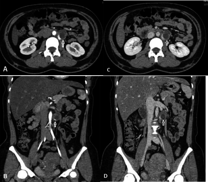
Retroperitoneal ectopic pregnancy is a rare form of ectopic pregnancy. In these cases, the gestational sac tends to attach near large retroperitoneal vessels, so when the sac ruptures, it can lead to severe bleeding, posing a life-threatening risk to the patient. Therefore, early diagnosis of ectopic pregnancy at this location is crucial. The diagnosis primarily relies on beta-hCG testing and imaging techniques, including ultrasound, computed tomography (CT), and magnetic resonance imaging (MRI). Our case is an ectopic pregnancy in the retroperitoneum that was diagnosed early through ultrasound and CT and successfully treated with surgery.

Keywords: CT scan; Ectopic; Pregnancy; Retroperitoneal; Ultrasound.


Figures

Fig 1
Fig. 1
(A) Ultrasound imaging did not show a gestational sac within the uterine cavity (UT: uterus, BL: bladder, Normal endometrial lining (asterisk); (B) Abdominal ultrasound imaging shows a ring-shaped mass in the retroperitoneal space (star shape), located adjacent to the left side of the abdominal aorta (AO).
Fig 2
Fig. 2
Arterial phase CT imaging (A, B) and venous phase CT imaging (C, D), the axial (A, C) and the sagittal (B, D) The imaging reveals a mass with a cystic component at the center, exhibiting marked contrast enhancement at the periphery (white arrow). The lesion is adjacent to the left side of the abdominal aorta and near the left gonadal vein.
Fig 3
Fig. 3
Pathologic examination reveals the presence of chorionic villi (black arrow) in the dissected tissue (hematoxylin-eosin stain, × 100).

References

    1. Maheux-Lacroix S., Li F., Bujold E., Nesbitt-Hawes E., Deans R., Abbott J. Cesarean scar pregnancies: a systematic review of treatment options. J Minimal Invas Gynecol. 2017;24(6):915–925. doi: 10.1016/j.jmig.2017.05.019. - DOI - PubMed
    1. Panelli D.M., Phillips C.H., Brady P.C. Incidence, diagnosis, and management of tubal and nontubal ectopic pregnancies: a review. Fertility Res Pract. 2015;1:15. doi: 10.1186/s40738-015-0008-z. - DOI - PMC - PubMed
    1. Xu H., Cheng D., Yang Q., Wang D. Multidisciplinary treatment of retroperitoneal ectopic pregnancy: a case report and literature review. BMC Pregnancy Childbirth. 2022;22(1):472. doi: 10.1186/s12884-022-04799-5. - DOI - PMC - PubMed
    1. Le D.N., Nguyen P.N., Huynh P.H. Retroperitoneal ectopic pregnancy: successful expectant management in condition of early pregnancy failure. BMC Pregnancy Childbirth. 2023;23(1):599. doi: 10.1186/s12884-023-05909-7. - DOI - PMC - PubMed
    1. Munzar Z., Afridi K.D., Amin F., Habib F. Retroperitoneal ectopic pregnancy presenting as a massive retroperitoneal hematoma. J College Physicians Surg–Pakistan. 2022;32(3):415–416. doi: 10.29271/jcpsp.2022.03.415. - DOI - PubMed

Publication types

LinkOut - more resources