Review
. 2025;2(1):18.
doi: 10.1038/s44341-025-00022-6. Epub 2025 Jul 4.

Mechanobiology in the eye

Affiliations
Review

Mechanobiology in the eye

C Ross Ethier et al. NPJ Biol Phys Mech. 2025.

Abstract

The eye presents a very dynamic biomechanical environment, and thus ocular cells must be highly mechanosensitive and mechanoresponsive. Moreover, defects in mechanobiological pathways contribute to a number of sight-threatening ocular diseases, highlighting the importance of ocular mechanobiology. We here give a concise overview of the mechanobiology of ocular cells in the lens and cornea (and how mechanobiology plays a role in associated pathologies in these tissues), before providing a detailed review of the mechanobiology of the common blinding disease, glaucoma. Mechanical stimuli are intimately linked with the pathology of glaucoma, both in terms of altered homeostasis of the eye's internal pressure control system and in the response of neural cells to elevated pressure in the eye. A complex array of mechanosensory elements (stretch-activated ion channels, integrins, G protein-coupled receptors) work together with intersecting networks of mechanotransducing pathways in cells of both the posterior and anterior eye in glaucoma. Despite intense research efforts over the past decades, much remains unknown about the mechanobiology of glaucoma. Continued investigation of glaucomatous mechanobiology is important, as it may reveal novel targets for treating this challenging disease.

Keywords: Atomic force microscopy; Diseases; Mechanisms of disease.


Conflict of interest statement

Competing interestsThe authors declare no competing interests.

Figures

Fig. 1
Fig. 1. Schematic overview of human ocular anatomy, showing the cornea, anterior chamber (AC), lens, vitreous, retina and sclera
. The inset at top right (red box) shows a magnified schematic view of the cornea, including (from anterior to posterior): the corneal epithelium (Epi), Bowman’s Layer (BL), the corneal stroma with resident keratocytes (dark blue), Descemet’s membrane (DM), and the corneal endothelium (Endo). The insets at top left show two magnified views of the conventional outflow pathway, located where the cornea, iris and sclera join. The purple boxed inset gives a schematic overview, while the green boxed image is a scanning electron micrograph from a human eye, in which Schlemm’s canal (SC), the trabecular meshwork (TM), the cornea (C) and the sclera (S) are identified. At lower right (orange box), the lens is shown, with details of its internal structure. Finally, at lower left (blue box), a schematic view of the optic nerve and surrounding structures is shown, with blood vessels and other structures identified, including the posterior ciliary arteries (PCA), the retina, the sclera (S), the central retinal artery and vein (CRA), the subarachnoid space, the optic nerve, the pia mater, the dural sheath, and the circle of Zinn and Haller (CZ). Immediately to its left (purple box) is an en face view of the optic nerve head region, in which a digestion process has been carried out to leave only the major connective tissues, namely the sclera and lamina cribrosa (LC). The schematic image of the cornea was created in Biorender.com and is based on ref. , the image of the lens is taken from ref. , the overview of the conventional outflow pathway is modified from ref. , the scanning electron micrograph of the outflow pathway is modified from ref. , the overview of the optic nerve head is modified from ref. , and the en face view of the optic nerve head is modified from ref. .
Fig. 2
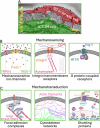
Fig. 2. The principal tissues of the outflow pathway and key mechanosensing and mechanotransduction pathways.
A Serial block-face scanning electron micrograph showing the interface between a Schlemm’s canal (SC) inner wall cell and a juxtacanalicular-trabecular meshwork (JCT-TM) cell, with direction of aqueous humor flow indicated by the yellow arrow (reproduced and adapted from ref. with permission from the authors under a Creative Commons license). B Schematic overview of relevant mechanosensing machineries including mechanosensitive ion channels (TRPV4, Piezo1, TREK-1), integrin transmembrane receptors, and G protein-coupled receptors (angiotensin II type 1 (AT1R) receptor). C Schematic overview of relevant mechanotransduction machineries including focal adhesion complexes (talin, paxillin, kindlin, vinculin, FAK, α-actinin, zyxin, VASP), cytoskeleton networks (actin filaments, intermediate filaments, microtubules), and shuttling proteins (YAP/TAZ, Wnt/β-catenin, glucocorticoid (GC)/soluble GC receptor). Schematics in (B, C) created using BioRender.com.
Fig. 3
Fig. 3. Overview of optic nerve head anatomy and relevant mechanobiologic components in glaucomatous optic neuropathy.
Black and white background shows detailed anatomy in a human eye (R = retina; S = sclera; LC = lamina cribrosa; CRV = central retinal vessel). Lower left inset shows individually labeled optic nerve head astrocytes in a cross-sectional view through the optic nerve at the level of the lamina cribrosa. Individual cell types are overlain on the background image (RGC = retinal ganglion cell, including myelin sheath posterior to the lamina cribrosa; A = optic nerve head astrocytes; MG = optic nerve head microglial cells). Note that cells are not to scale. Inset at upper left show mechanobiologic machinery in these cell types. MSIC = mechanosensitive ion channel, including TRPV4 and Piezo1; MAPKs = mitogen-activated protein kinases, including ERK, c-JUN/JNK, p38 and p42/44. Background image from ref. with permission; lower left inset from ref. , with permission. Cell overlays and mechanobiologic insets created using BioRender.com.

References

    1. Brown, M. M., Brown, G. C., Sharma, S. & Busbee, B. Quality of life associated with visual loss: a time tradeoff utility analysis comparison with medical health states. Ophthalmology110, 1076–1081 (2003). - PubMed
    1. Vu, H. T., Keeffe, J. E., McCarty, C. A. & Taylor, H. R. Impact of unilateral and bilateral vision loss on quality of life. Br. J. Ophthalmol.89, 360–363 (2005). - PMC - PubMed
    1. Chen, Y., Ju, L., Rushdi, M., Ge, C. & Zhu, C. Receptor-mediated cell mechanosensing. Mol. Biol. Cell28, 3134–3155 (2017). - PMC - PubMed
    1. Martino, F., Perestrelo, A. R., Vinarský, V., Pagliari, S. & Forte, G. Cellular Mechanotransduction: from tension to function. Front. Physiol.9, 824 (2018). - PMC - PubMed
    1. Grytz, R., Yang, H., Hua, Y., Samuels, B. C. & Sigal, I. A. Connective tissue remodeling in myopia and its potential role in increasing risk of glaucoma. Curr. Opin. Biomed. Eng.15, 40–50 (2020). - PMC - PubMed

LinkOut - more resources