

Diagnostic Pitfalls in Peritoneal Carcinomatosis: A Case of Pseudomyxoma Peritonei
- PMID: 40621304
- PMCID: PMC12229253
- DOI: 10.7759/cureus.85474
Diagnostic Pitfalls in Peritoneal Carcinomatosis: A Case of Pseudomyxoma Peritonei
Abstract
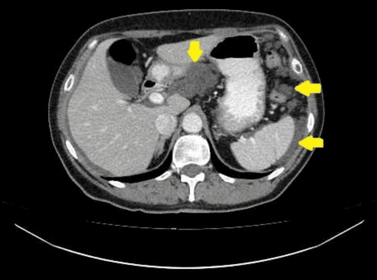
Pseudomyxoma peritonei (PMP) is a rare pathological condition posing significant diagnostic and management challenges. This article presents a clinical case of a 58-year-old female who was initially diagnosed with stage III primary peritoneal carcinoma. Following neoadjuvant chemotherapy, a diagnostic re-evaluation was performed with histopathological and immunohistochemical review of biopsy specimens, which led to a revised diagnosis of mucinous carcinoma with features consistent with PMP. The patient subsequently underwent complete cytoreductive surgery (CRS) followed by hyperthermic intraperitoneal chemotherapy (HIPEC). She has remained disease-free for 12 months post-operatively. This case illustrates the crucial role of pathological assessment in guiding treatment for PMP and demonstrates favorable long-term outcomes with aggressive CRS and HIPEC in appropriately selected patients.
Keywords: cytoreductive surgery; gynecologic oncology; hyperthermic intraperitoneal chemotherapy; mucinous neoplasms; pseudomyxoma peritonei.
Copyright © 2025, Isachanka et al.
Conflict of interest statement
Human subjects: Consent for treatment and open access publication was obtained or waived by all participants in this study. N. N. Alexandrov National Cancer Centre of Belarus issued approval Not Applicable. As per institutional policy overseen by the N.N. Alexandrov National Cancer Centre of Belarus, formal ethics approval is waived for single case reports where written informed consent for publication has been obtained from the patient. Conflicts of interest: In compliance with the ICMJE uniform disclosure form, all authors declare the following: Payment/services info: All authors have declared that no financial support was received from any organization for the submitted work. Financial relationships: All authors have declared that they have no financial relationships at present or within the previous three years with any organizations that might have an interest in the submitted work. Other relationships: All authors have declared that there are no other relationships or activities that could appear to have influenced the submitted work.
Figures


References
-
- Uber das sogennante Pseudomyxoma Peritonei. (Article in Portuguese) Frankel E. https://scholar.google.com/scholar_lookup?title=Uber%20das%20sogenannte%... Munch Med Wschr. 1901;48:965–970.
-
- Results of treatment of 385 patients with peritoneal surface spread of appendiceal malignancy. Sugarbaker PH, Chang D. Ann Surg Oncol. 1999;6:727–731. - PubMed
Publication types
LinkOut - more resources
Full Text Sources