. 2025 Jul 16;17(28):40237-40257.
doi: 10.1021/acsami.5c08237. Epub 2025 Jul 7.

Long-Gap Sciatic Nerve Regeneration Using 3D-Printed Nerve Conduits with Controlled FGF-2 Release

Affiliations

Long-Gap Sciatic Nerve Regeneration Using 3D-Printed Nerve Conduits with Controlled FGF-2 Release

Diego N Rodriguez-Sanchez et al. ACS Appl Mater Interfaces. .

Abstract

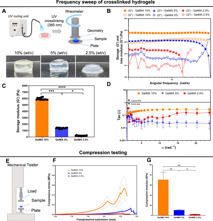
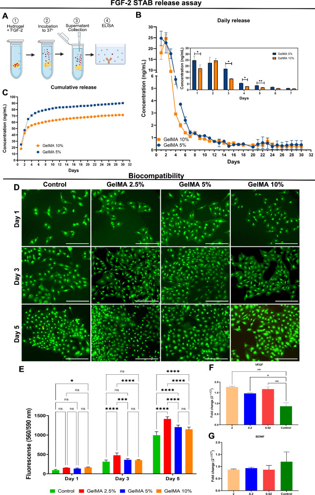
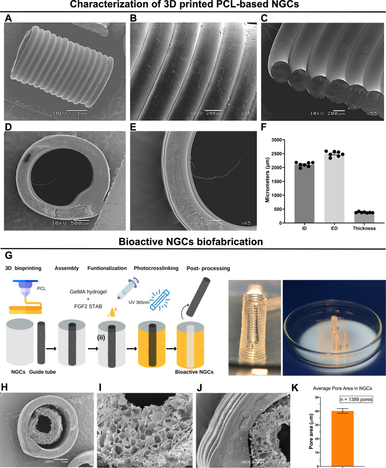
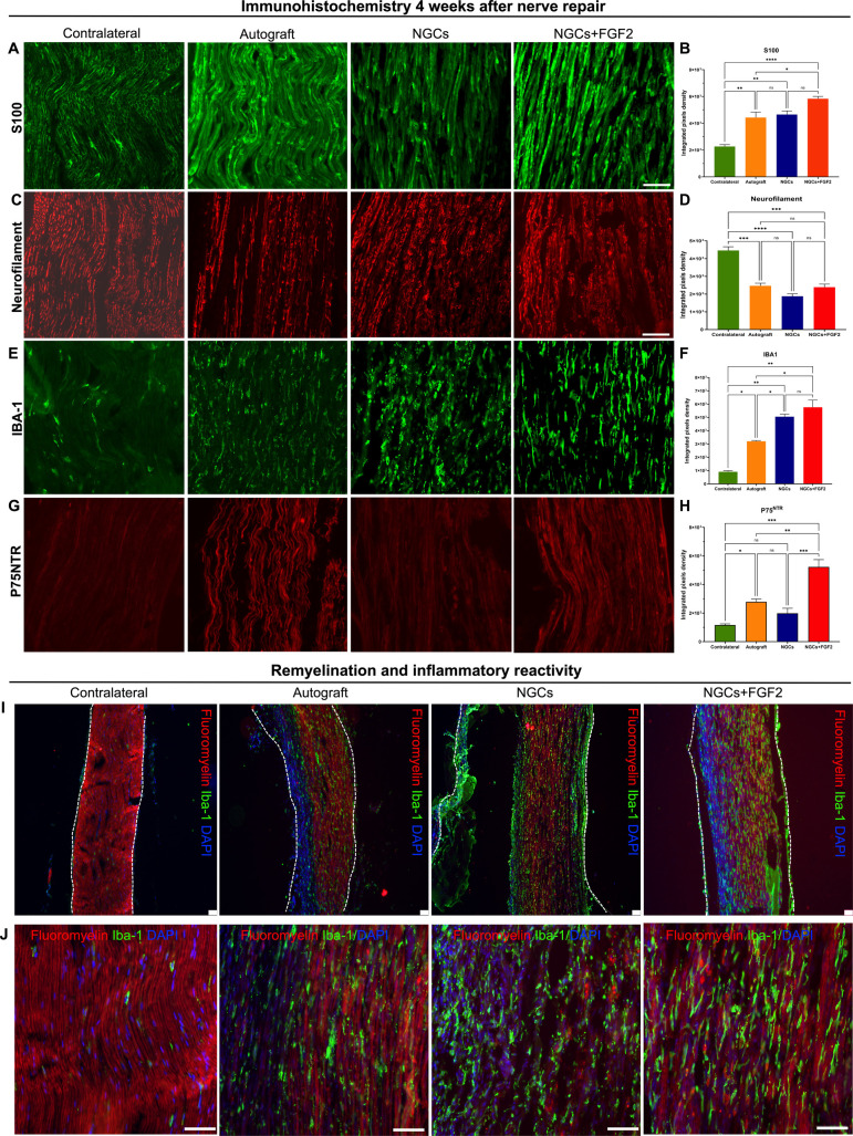
Peripheral nerve injuries (PNIs) by transection require reconstructive surgery, often with highly variable results and persistent sensory and motor deficits. Three-dimensional (3D) printing enables the biofabrication of nerve guidance conduits (NGCs) with the ability to release neurotrophic factors, showing therapeutic potential. We developed a 3D printing process of NGCs using polycaprolactone (PCL) and gelatin methacryloyl (GelMA) integrated with a thermostable fibroblast growth factor 2 (FGF-2). The synthesized GelMA at 10% (w/v) concentration showed superior rheological, mechanical, and ultrastructural characteristics, ensuring 3D printing fidelity. Incorporating FGF-2 into GelMA resulted in a controlled release pattern over 30 days along with biocompatibility and an increase of metabolism in rat S16 Schwann cells and human mesenchymal stem cells (MSCs). MSCs exhibited gene regulation linked to vascularization after FGF-2 stimulation. The PCL polymer facilitated the buildability of a spiral-patterned tubular structure, which was functionalized with a combination of GelMA and UV photocrosslinked. At 12 weeks, following a long-gap nerve injury in rats, NGC implantation enhanced sensory and motor recovery, improved electrophysiological function, and promoted morphological and ultrastructural nerve reorganization and regeneration. At 4 weeks, significant Schwann cell proliferation (S100), expression of the pan-neurotrophin receptor (P75NTR), myelination of newly grown axons, and organization of neurofilaments were observed. The bioactive NGCs represent a promising alternative to nerve autografts for the repair of long-gap injuries.

Keywords: 3D printing; FGF-2; biomaterials; gelatin methacryloyl; nerve guidance conduits; peripheral nerve regeneration; polycaprolactone.


Figures

1
1
Rheological and ultrastructural characteristics of the hydrogel 2.5%, 5%, and 10% GelMA. (A) Viscosity as a function of the temperature. (B) Viscosity as a function of the shear rate at 22 °C. (C) Sweep temperature on G′ and G″. All analyses: n = 2. Black arrows indicate the gel points of each concentration. SEM characterization of GelMA constructs after UV photo-cross-linking of (D) 5% and (E) 10% (w/v) GelMA 24 h postlyophilization at 130× and 700× magnification, respectively. Scale bars: 20 and 100 μm. Values are represented as mean ± SEM. *p < 0.0332, **p < 0.0021, ***p < 0.0002, and ****p < 0.0001.
2
2
Frequency sweep analysis and compression testing of the hydrogel compositions. (A) Schematic diagram indicating how the samples were subjected to UV photocrosslinking (240 s, 18 mW cm–2) to obtain cross-linked 10%, 5%, and 2.5% GelMA hydrogels following of rotational rheometry. (B) G′ and G″ as a function of the angular frequency (ω). (C) Comparison of G′ between the groups. (D) Damping factor (tan δ) as a function of ω. All analyses: n = 2. (E) Compression test using a mechanical Instron. (F) Stress and extension curve from cross-linked hydrogels. (G) Compressive modulus mean values from cross-linked hydrogels (n = 3). Values are represented as mean ± SEM. *p < 0.0332, **p < 0.0021, ***p < 0.0002, and ****p < 0.0001.
3
3
Quantification of FGF-2 release and cytotoxicity assessment in S16 cells. (A) Schematic diagram summarizing the FGF-2 release assay over 30 days. (B) Daily release evidenced a burst release in the first week. The inset in panel B compares the FGF-2 releases between 5% and 10% GelMA during the initial 7 days. (C) Cumulative release of FGF-2 over 30 days. (D) Representative images of S16 cells following indirect contact with GelMA+FGF-2, showing live cells (green, calcein AM) and dead cells (red, ethidium homodimer). (E) Presto Blue quantification in 1, 3, and 5 days following indirect exposure with GelMA+FGF-2. Scale bar: 300 μm. Relative gene expression of VEGF (F) and BDNF (G) after 3 days of stimulation of MSCs with FGF-2. All analyses: n = 3. Amplification of GAPDH was used as the endogenous gene. Data are presented as mean ± SEM. *p < 0.0332, **p < 0.0021, ***p < 0.0002, and ****p < 0.0001.
4
4
Structural characterization and fabrication process of 3D-printed NGCs. (A) Top, (B) dorsal, and (C) fractured lateral views of the 3D-printed NGCs featuring a spiral pattern and distinct individual filament structures. Cross-sectional views at midlevel of the NGCs at low (D) and high (E) magnification. (F) Analysis of the dimensions of NGCs including the inner diameter, external diameter, and wall thickness. Scale bars: 1 mm and 200 and 500 μm. (G) Schematic diagram illustrating the procedures involved in the biofabrication of NGCs and their macroscopic appearance. (H) Top view of the NGCs following cross-linking of the internal 10% GelMA layer incorporated with FGF-2. (I) Detailed view showing preservation of the NGC lumen after removal of the guide tube. (J) Magnified view of the interface between PCL and GelMA. (K) Analysis of the average pore size on the internal wall of 10% GelMA based on SEM images (n = 3). Scale bars: 200 μm and 1 mm. Data are presented as mean ± SEM.
5
5
Experimental sciatic nerve injury in the autograft, NCGs, and NGCs+FGF2 groups. (A) Schematic diagram demonstrating how the regenerative process is irreversible after long-gap PNI lesions. (B) Bioactive NGCs fabricated by using 3D printing and implanted into an 8-mm long-gap PNI rat model for subsequent functional and morphological evaluation. (C) In the autograft group, sciatic nerve neurotmesis (8 mm critical defect) performed, repositioned, and repaired with epineurial sutures. In the NGCs and NGCs+FGF2 groups, critical defect of the sciatic nerve was created, and then the nerve stumps were inserted 1 mm into the NGCs and fixed by epineural sutures. Scale bar: 10 mm.
6
6
Analysis of the locomotor, sensorial, and electrophysiological recovery for 12 weeks after nerve repair. (A) Schematic illustrations of the catwalk platform system and the electronic von Frey system. Comparisons of the peroneal functional index (B), contact area (C), maximum contact area (D), base of support for hindlimbs (E) and forelimbs (F), regularity index (G), and withdrawal threshold (Δ withdrawal thresholds) (H) across the experimental groups. (I) Electrophysiological procedure to obtain an CMAP from the tibialis cranialis muscle. (J) Nerve conduction velocity obtained after proximal and distal stimulation. Measurements included (K) latency, (L) amplitude, and (M) duration in the experimental groups at 12 weeks. All analyses: n = 5. The values are represented as mean ± SEM. *p < 0.0332, **p < 0.0021, ***p < 0.0002, and ****p < 0.0001.
7
7
Analysis of neuromuscular reinnervation 12 weeks postnerve repair. (A) Microscopic comparison of the lesioned and contralateral tibialis cranialis and gastrocnemius muscles across the experimental groups. Graphs show comparative data for gastrocnemius muscle weight (B), tibialis cranialis muscle weight (C), and overall body weight (D) among the autograft, NGCs, and NGCs+FGF2 groups. All analyses: n = 5. The values are represented as mean ± SEM. *p < 0.0332, **p < 0.0021, ***p < 0.0002, and ****p < 0.0001.
8
8
Analysis of immunostaining for cytoskeletal components, neurotrophin receptors, degree of myelination, and inflammatory reactivity 30 days postinjury in autografts, NGCs, and NGCs with FGF-2 groups. Immunostaining included S100 (A), neurofilament (C), IBA-1 (E), and P75NTR (G), with corresponding quantification of the integrated pixel density shown in parts B, D, F, and H, respectively. Representative low-magnification images (I) at 10× demonstrate myelin sheath labeling with fluoromyelin (red) and macrophages with IBA-1 (green), indicating reinnervation of proximal and distal nerve stumps. High-magnification images (J) at 40× show triple labeling with IBA-1 (green), DAPI (blue), and fluoroyelin (red). All analyses: n = 3. The values are represented as mean ± SEM. *p < 0.0332, **p < 0.0021, ***p < 0.0002, and ****p < 0.0001.
9
9
Ultrastructural and morphometric analysis of the myelinated fibers, axon diameters, myelin sheath thicknesses, and g ratios was performed at 12 weeks following PNI and repair in the experimental groups. (A) High-resolution TEM images showing the organization and distribution of myelinated axons, myelin sheath thicknesses, and endoneural collagens across the groups at 12 weeks. Measurements of the fiber diameter (B), axon diameter (C), myelin sheath thickness (D), and g ratio (E) were obtained from motor axons and compared among the contralateral, autograft, NGCs, and NGCs + FGF2 groups. The insets in part E present the correlation analysis between the g ratio and axon diameter (μm). All analyses: n = 5. Scale bars: 1 and 5 μm.

References

    1. Taylor C. A., Braza D., Rice J. B., Dillingham T.. The Incidence of Peripheral Nerve Injury in Extremity Trauma. Am. J. Phys. Med. Rehabil. 2008;87(5):381–385. doi: 10.1097/PHM.0b013e31815e6370. - DOI - PubMed
    1. Karsy M., Watkins R., Jensen M. R., Guan J., Brock A. A., Mahan M. A.. Trends and Cost Analysis of Upper Extremity Nerve Injury Using the National (Nationwide) Inpatient Sample. World Neurosurg. 2019;123:e488–e500. doi: 10.1016/j.wneu.2018.11.192. - DOI - PubMed
    1. Huckhagel T., Nüchtern J., Regelsberger J., Lefering R.. Nerve Injury in Severe Trauma with Upper Extremity Involvement: Evaluation of 49,382 Patients from the TraumaRegister DGU® between 2002 and 2015. Scand. J. Trauma. Resusc. Emerg. Med. 2018;26(1):76. doi: 10.1186/s13049-018-0546-6. - DOI - PMC - PubMed
    1. Bergmeister K. D., Große-Hartlage L., Daeschler S. C., Rhodius P., Böcker A., Beyersdorff M., Kern A. O., Kneser U., Harhaus L.. Acute and Long-Term Costs of 268 Peripheral Nerve Injuries in the Upper Extremity. PLoS One. 2020;15(4):1–12. doi: 10.1371/journal.pone.0229530. - DOI - PMC - PubMed
    1. Burrell J. C., Ali Z. S., Zager E. L., Rosen J. M., Tatarchuk M. M., Cullen D. K.. Engineering the Future of Restorative Clinical Peripheral Nerve Surgery. Adv. Healthc. Mater. 2025;2404293:1–24. doi: 10.1002/adhm.202404293. - DOI - PubMed

MeSH terms