. 2025 Jul 1;18(7):dmm052258.
doi: 10.1242/dmm.052258. Epub 2025 Jul 28.

Distinguishing PEX2 and PEX16 gene variant severity for mild, severe and atypical peroxisome biogenesis disorders

Affiliations

Distinguishing PEX2 and PEX16 gene variant severity for mild, severe and atypical peroxisome biogenesis disorders

Vanessa A Gomez et al. Dis Model Mech. .

Abstract

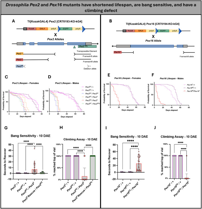
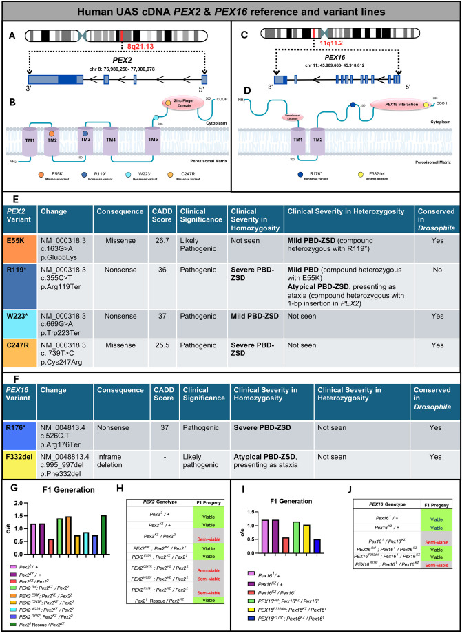
Peroxisomal biogenesis disorders (PBD) are autosomal recessive diseases caused by mutations in specific PEX genes that impair peroxisome formation, leading to multi-systemic failure. Symptoms vary, even in patients with variants in the same PEX gene. Our goal is to select PEX mutations and use Drosophila to model a severity spectrum based on genotype-phenotype correlations. Utilizing KozakGAL4 (KZ) cassettes, we replaced the coding sequence of Pex with a GAL4 driver, ideal for making 'humanized' flies in which human PEX can replace the fly loss. We generated Pex2KZ and Pex16KZ lines and assessed them in various behavior assays, confirming their severe phenotypes. We performed rescue with human reference, variant PEX2 and PEX16 alleles, and phenotypic rescue was observed when human PEX2Ref or PEX16Ref were expressed in Pex2KZ or Pex16KZ flies, respectively. We identified a severity spectrum for PEX2 and PEX16 alleles, with some missense mutations exhibiting severity comparable to truncations. Alleles linked to mild PBD showed partial rescue, while variants associated with atypical ataxia could fully rescue. Drosophila humanization is an effective method to study the range of severity of PBD.

Keywords: Drosophila; PBD; PEX16; PEX2; Peroxisome; Zellweger.


Conflict of interest statement

Competing interests The authors declare no competing or financial interests.

Figures

Fig. 1.
Fig. 1.
The KozakGAL4 knock-in/knock-out strategy, which replaces the coding region of the Pex gene of interest by identifying sgRNA target sites within the 5′ UTR and 3′ UTR. (A) Schematics of Pex2 locus and targeting strategy. (B) Schematics of Pex16 locus and targeting strategy. Gray boxes in A and B indicate UTRs; blue boxes indicate the Pex-coding region. (C-C″) Immunofluorescence images showing the Pex2KZ expression pattern in third-instar Drosophila larval brains. Pex2KZ expression (mCherry, blue) is shown in C. Staining against Repo (nuclear glia cells, purple) or Elay (neurons, green) is shown in C′ or C″, respectively. (C‴) Merged image showing colocalization within the ventral nerve cord, indicating Pex2KZ (blue) expression in both neurons (green) and nuclear glia (purple) of the larval brain. (D-D″) Immunofluorescence images showing the Pex16KZ expression pattern in third-instar Drosophila larval brains. Pex16KZ expression (mCherry, blue) is shown in D. Staining against Repo (nuclear glia cells, purple) or Elav (neurons, green) is shown in D′ or D″, respectively. (D‴) Merged image showing colocalization within the ventral nerve cord, indicating Pex16KZ (blue) expression in both neurons (green) and nuclear glia (purple) in the larval brain. Scale bars: 100 µm.
Fig. 2.
Fig. 2.
Drosophila Pex2 and Pex16 mutants have shortened lifespan, are bang sensitive and have a climbing defect. (A) Schematic representation of the fly Pex2 gene along with four alleles, including a coding P-element insertion (Pex2EPg), two deletion alleles (Pex21, Pex22) and Pex2-KozakGAL4 (Pex2KZ). (B) Schematic representation of the fly Pex16 gene along with one frameshift alleles (Pex161) and Pex16-KozakGAL4 (Pex16KZ). (C) Pex2 female lifespan assay shows that the Pex2 mutants have a shorter lifespan compared to Pex2 control lines (n=51 Pex2Df/+ and n=50 Pex2KZ/+). n=55 Pex2KZ/Pex2Df, n=54 Pex2KZ/Pex21, n=58 Pex2KZ/Pex22, n=57 Pex2KZ/Pex2EPg. (D) Pex2 male lifespan assay shows that the Pex2 mutants have a shorter lifespan compared to Pex2 control lines (n=37 Pex2Df/+ and n=48 Pex2KZ/+). n=56 Pex2KZ/Pex2Df, n=55 Pex2KZ/Pex21, n=57 Pex2KZ/Pex22, n=53 Pex2KZ/Pex2EPg. (E) Pex16 female lifespan assay shows that the Pex16KZ/Pex161 mutant flies (n=51) have a shorter lifespan compared to Pex16 control lines (n=48 Pex161/+ and n=50 Pex16KZ/+). (F) Pex16 male lifespan assay shows that the Pex16KZ/Pex161 mutant flies (n=51) have a shorter lifespan compared to Pex16 control lines (n=50 Pex161/+ and n=50 Pex16KZ/+). (G) Pex2 null flies have a significant bang-sensitive phenotype (Pex2KZ/Pex22, n=36) compared to Pex2 control lines (n=40 Pex22/+ and n=40 Pex2KZ/+) and Pex22 rescue (n=29) observed at 10 days after eclosion (DAE). (H) Pex2 null flies have a significant climbing deficiency (Pex2KZ/Pex22, n=36) compared to Pex2 control lines (n=40 Pex22/+ and n=40 Pex2KZ/+) and Pex22 rescue (n=29) observed at 10 DAE. (I) Pex16 null flies have a significant bang-sensitive phenotype (Pex16KZ/Pex161, red n=32) compared to Pex16 control lines (n=40 Pex161/+ and n=40 Pex16KZ/+) observed at 10 DAE. (J) Pex16 null flies have a significant climbing deficiency (Pex161/Pex16KZ, n=32) compared to Pex16 control lines (n=40 Pex161/+ and n=40 Pex16KZ/+) observed at 10 DAE. ***P<0.001, ****P<0.0001.
Fig. 3.
Fig. 3.
Human UAS cDNA PEX2 and PEX16 reference and variant lines. (A) Schematic representation of human PEX2 gene. (B) Schematic representation of human PEX2 protein and variant locations. (C) Schematic representation of human PEX16 gene. (D) Schematic representation of human PEX16 protein and variant locations. (E) PEX2 variant table indicates the consequence of the change, pathogenicity prediction, clinical significance, clinical severity in homozygosity and heterozygosity, and conservation in Drosophila. (F) PEX16 variant table indicates the consequence of the change, pathogenicity prediction, clinical significance, clinical severity in homozygosity and heterozygosity, and conservation in Drosophila. (G) Plotted are the observed/expected Mendelian ratios of the F1 generation of human PEX2 variants, in a fly null background. n=306 Pex22/+, n=345 Pex2KZ/+, n=273 Pex2KZ/Pex22, n=214 PEX2Ref, n=135 PEX2E55K, n=172 PEX2C247R, n=365 PEX2W223*, n=337 PEX2R119*, n=136 Pex22 Rescue/Pex2KZ. (H) Assessment of the PEX2 phenotype and its F1 progenies identified as lethal, viable, or semi-viable. (I) Plotted are the observed/expected Mendelian ratios of the F1 generation of human PEX16 variants, in a fly null background. n=292 Pex161/+, n=270 Pex16KZ/+, n=126 Pex16KZ/Pex161, 278 PEX16Ref, 197 PEX16F332del, 214 PEX16R176*. (J) Assessment of the PEX16 phenotype and its F1 progenies identified as lethal, viable or semi viable. For the calculation of the observed/expected Mendelian ratios of the F1 generations see Table S3.
Fig. 4.
Fig. 4.
Pex3 immunostaining in third-instar larva body wall muscle. (I, top) Images show control group flies (Pex2KZ/+) (A-A″), Pex2 null flies (Pex2KZ/Pex22), (B-B″) and human rescue group flies (PEX2Ref;Pex2KZ/Pex22) (C-C″). Pex3-positive puncta are shown in red; nuclei were stained with DAPI (blue). Merged images are shown in A″-B″. (D) Quantification and comparison of flies shown in A-C″ with Pex3-positive puncta comprising an area >0.5µm2. n=7 Pex2KZ/+ (control), n=7 Pex2KZ/Pex22, n=6 PEX2Ref;Pex2KZ/Pex22. (II, bottom) Images show control group flies (Pex16KZ/+) (A-A″), Pex16 null flies (Pex16KZ/Pex162) (B-B″) and human rescue group flies (PEX16Ref;Pex16KZ/Pex162) (C-C″). Pex3-positive puncta are shown in red; nuclei were stained with DAPI (blue). Merged images are shown in A″-B″. (D) Quantification and comparison of flies shown in A-C″ with Pex3-positive puncta comprising an area >0.5 µm2 between the three genotypes. n=7 Pex16KZ/+ control, n=7 Pex16KZ/Pex161, n=7 PEX16Ref. *P<0.05, **P=0.0011, ***P=0.001, ****P<0.0001. All scale bars: 10 µm
Fig. 5.
Fig. 5.
Rescue-based humanization of Pex2 and Pex16 behavior assays. (A) Lifespan analysis of PEX2Ref and Variant flies (n=110 PEX2Ref, n=98 PEX2E55K, n=101 PEX2C247R, n=102 PEX2W223*, n=101 PEX2R119*) along with our Pex2 null (Pex2KZ/Pex22 n=115) and Pex2 control lines (n=117 Pex22/+ and n=98 Pex2KZ/+). (B) Bang-sensitivity assay at 10 days after eclosion (DAE) of PEX2Ref and Variant flies (n=26 PEX2Ref, n=37 PEX2E55K, n=38 PEX2C247R, n=32 PEX2W223*, n=34 PEX2R119*), along with Pex2KZ/Pex22 (n=36) and Pex2 control lines (n=40 Pex22/+ and n=40 Pex2KZ/+). (C) Bang-sensitivity assay of PEX2Ref and Variant flies at 15 DAE (n=26 PEX2Ref, n=32 PEX2E55K, n=32 PEX2C247R, n=22 PEX2W223*, n=31 PEX2R119*), along with Pex2KZ/Pex22 (n=23 and Pex2 control lines (n=40 Pex22/+ and n=40 Pex2KZ/+). (D) Climbing assay at 5 DAE of PEX2Ref and Variant flies with data indicating if flies either had the ability to climb or not (n=30 PEX2Ref, n=40 PEX2E55K, n=40 PEX2C247R, n=40 PEX2W223*, n=40 PEX2R119*), along with Pex2KZ/Pex22 (n=45) and Pex2 control lines (n=40 Pex22/+ and n=40 Pex2KZ/+). (E) Climbing assay at 10 DAE of PEX2Ref and Variant flies (n=26 PEX2Ref, n=37 PEX2E55K, n=38 PEX2C247R, n=32 PEX2W223*, n=34 PEX2R119*), along with Pex2KZ/Pex22 (n=36) and Pex2 control lines (n=40 Pex22/+ and n=40 Pex2KZ/+). (F) Climbing assay at 15 DAE of PEX2Ref and Variant flies (n=26 PEX2Ref, n=32 PEX2E55K, n=32 PEX2C247R, n=22 PEX2W223*, n=31 PEX2R119*), along with Pex2KZ/Pex22 (n=23) and Pex2 control lines (n=40 Pex22/+ and n=40 Pex2KZ/+). (G) Lifespan analysis of PEX16Ref and Variant flies (n=100 PEX16Ref, n=100 PEX16F332del, n=98 PEX16R176*), along with our Pex16 null Pex16KZ/Pex161 (n=102) and Pex16 control lines (n=100 Pex161/+ and n=100 Pex16KZ/+). (H) Bang-sensitivity assay at 10 DAE of PEX16Ref and Variant flies (n=35 PEX16Ref, n=40 PEX16F332del, n=31 PEX16R176*), along with Pex16KZ/Pex161 n=32 and Pex16 control lines (n=40 Pex161/+ and n=40 Pex16KZ/+). (I) Bang-sensitivity assay at 15 DAE of PEX16Ref and Variant flies (n=30 PEX16Ref, n=38 PEX16F332del, n=23 PEX16R176*), along with Pex16KZ/Pex161 (n=23) and Pex16 control lines (n=40 Pex161/+ and n=40 Pex16KZ/+). (J) Climbing assay at 5 DAE of PEX16Ref and Variant flies with data indicating if flies either had the ability to climb or not (n=40 PEX16Ref, n=40 PEX16F332del, n=40 PEX16R176*), along with Pex16KZ/Pex161 (n=40) and Pex16 control lines (n=40 Pex161/+ and n=40 Pex16KZ/+). (K) Climbing assay at 10 DAE of PEX16Ref and Variant flies (n=35 PEX16Ref, n=40 PEX16F332del, n=31 PEX16R176*), along with Pex16KZ/Pex161 (n=32) and Pex16 control lines (n=40 Pex161/+ and n=40 Pex16KZ/+). (L) Climbing assay at 15 DAE of PEX16Ref and Variant flies (n=30 PEX16Ref, n=38 PEX16F332del, n=23 PEX16R176*), along with Pex16KZ/Pex161 (n=23) and Pex16 control lines (n=40 Pex161/+ and n=40 Pex16KZ/+). *P<0.05, **P<0.01, ***P<0.001, ****P<0.0001; ns, not significant.
Fig. 6.
Fig. 6.
Thickening of the motor axon within DFM49 of adult flies. (I, top) Panels A-A‴ represent Pex2 control group flies (Pex2KZ/+); panels B-B‴ represent Pex2 null flies (Pex2KZ/Pex22) and panels C-C‴ represent human PEX2 rescue group flies (PEX2Ref;Pex2KZ/Pex22). Dashed lined surround the DFM49. Panels A-C show staining for peroxisomes (Pex3, purple), panels A′-C′ show staining for nerve fiber tracks (HRP, green). Panels A″-C″ show nuclei (blue) stained with DAPI. Boxed areas in A-C are shown magnified in A″″-C″″, respectively. (D) Schematic representation of the DFM49, with numbers within indicating the motor axon entering the flight muscle (1), the thick branch (2a) and the thin branch (2b). (E) Quantification of the Pex2 DFM49 motor axon width between the three genotypes. (F) Quantification of the thick branch of the Pex2 DFM49 motor axon width between the three genotypes. (G) Quantification of the thin branch of the Pex2 DFM49 motor axon width between the three genotypes. n=4 Pex2KZ/+ control, n=4 Pex2KZ/Pex22, n=4 PEX2Ref. (II, bottom) Panels A-A‴ represent Pex16 control group flies (Pex16KZ/+); panels B-B‴ represent Pex16 null flies (Pex16KZ/Pex162) and panels C-C‴ represent human PEX16 rescue group flies (PEX16Ref;Pex16KZ/Pex162). Dashed lined surround the DFM49. Panels A-C show staining for peroxisomes (Pex3, purple), panels A′-C′ show staining for nerve fiber tracts (HRP, green). Panels A″-C″ show nuclei (blue) stained with DAPI. Boxed areas in A-C are shown magnified in  A″″-C″″, respectively. (D) Schematic representation of the DFM49, with numbers within indicating the motor axon entering the flight muscle (1), the thick branch (2a) and the thin branch (2b). (E) Quantification of Pex16 DFM49 motor axon width between the three genotypes. (F) Quantification of the thick branch of the Pex16 DFM49 motor axon width between the three genotypes. (G) Quantification of the thin branch of the Pex16 DFM49 motor axon width between the three genotypes. n=4 Pex16KZ/+ control, n=4 Pex16KZ/Pex161, n=4 PEX16Ref. ns, not significant. *P<0.05, **P<0.01, ***P<0.001. Scale bars: 20 µm (A-C‴ in I and II), 5 µm (A″″-C″″ in I and II).

Update of

Similar articles

References

    1. Bacino, C., Chao, Y.-H., Seto, E., Lotze, T., Xia, F., Jones, R. O., Moser, A. and Wangler, M. F. (2015a). Dataset for a case report of a homozygous PEX16 F332del mutation. Data Brief 6, 722-727. 10.1016/j.dib.2015.12.011 - DOI - PMC - PubMed
    1. Bacino, C. A., Chao, Y.-H., Seto, E., Lotze, T., Xia, F., Jones, R. O., Moser, A. and Wangler, M. F. (2015b). A homozygous mutation in PEX16 identified by whole-exome sequencing ending a diagnostic odyssey. Mol. Genet. Metab. Rep. 5, 15-18. 10.1016/j.ymgmr.2015.09.001 - DOI - PMC - PubMed
    1. Baumgartner, M. R. and Saudubray, J. M. (2002). Peroxisomal disorders. Semin. Neonatol. 7, 85-94. 10.1053/siny.2001.0089 - DOI - PubMed
    1. Bellen, H. J. and Yamamoto, S. (2015). Morgan's legacy: fruit flies and the functional annotation of conserved genes. Cell 163, 12-14. 10.1016/j.cell.2015.09.009 - DOI - PMC - PubMed
    1. Bose, M., Yergeau, C., D'Souza, Y., Cuthbertson, D. D., Lopez, M. J., Smolen, A. K. and Braverman, N. E. (2022). Characterization of severity in Zellweger spectrum disorder by clinical findings: a scoping review, meta-analysis and medical chart review. Cells 11, 1891. 10.3390/cells11121891 - DOI - PMC - PubMed

Substances

Supplementary concepts

LinkOut - more resources