Review
. 2025 Jul 15;39(13):e70810.
doi: 10.1096/fj.202500737R.

Food Additives: Emerging Detrimental Roles on Gut Health

Affiliations
Review

Food Additives: Emerging Detrimental Roles on Gut Health

Tyler Seto et al. FASEB J. .

Abstract

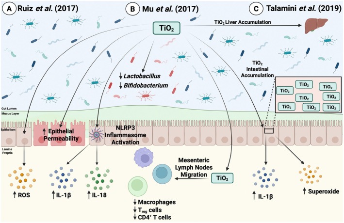
Processed and ultra-processed foods have become dietary staples in many developed countries. A major constituent of these foods is a variety of synthetic chemical additives, which are used to improve the texture, preservation, and aesthetics of food. Evidence is mounting that synthetic chemicals used as food additives may have harmful impacts on health. Studies have linked certain additives to health conditions such as attention deficit hyperactivity disorder, cancer, and obesity. In addition, emerging evidence suggests that additives, such as emulsifiers, artificial sweeteners, colorants, and preservatives, may act as potential disruptors of intestinal homeostasis. Indeed, various studies have identified that food additives can impact gut health by modulating gut microbiota and intensifying intestinal inflammation. Considering the lack of known nutritional benefits of these additives and the accumulating evidence on the detrimental effects of these additives on gut health, further experimental, epidemiological, and clinical evaluations are imperative. This will provide significant advances in the prevention and management of gut health, including intestinal inflammation, and in enriching public knowledge on the harmful effects of these additives. In this review, we explore the effects of popular food additives on gut health with a particular focus on intestinal inflammation and examine the broader implications of these impacts on food safety policy and public health.

Keywords: food additives; food dye; food emulsifier; gut health; intestinal inflammation; microbiota; processed/ultra‐processed foods; public health.


Conflict of interest statement

The authors declare no conflicts of interest.

Figures

FIGURE 1
FIGURE 1
The effects of various azo dyes on GI health. (A) He et al. reported that azo reduction of Allura Red (AR) and Sunset Yellow (SY) by B. ovatus and E. faecalis produced the metabolite ANSA‐Na, leading to an IL‐23‐dependent colitis mediated by CD4+ T‐cells and IFN‐γ production [23]. (B) Kwon et al. identified that chronic AR exposure promoted experimental colitis via colonic 5‐HT in both a gut microbiota‐dependent and ‐independent pathway and by decreasing intestinal barrier integrity through MLCK [24]. (C) Zahran and colleagues showed that SY exposure activated the NLRP3 inflammasome, leading to the production of IL‐18 and IL‐1β and inducing microbial dysbiosis, which resulted in increased LPS production and impaired barrier function via altered adherens junction (AJ) complexes [25]. Image created in BioRender.
FIGURE 2
FIGURE 2
Effects of TiO2 on intestinal homeostasis. (A) Ruiz et al. identified that titanium dioxide (TiO2) administration exacerbated DSS‐induced colitis through increased reactive oxygen species (ROS) production, increased intestinal epithelial permeability, and activated the NLRP3 inflammasome, which subsequently led to increased pro‐inflammatory cytokine (IL‐1β and IL‐18) production [34]. (B) Mu and colleagues showed that TiO2 exposure altered the microbial composition by decreasing Bifidobacterium and Lactobacillus abundance. TiO2 also migrated to the mesenteric lymph nodes and reduced CD4+ T‐cells, regulatory T‐cells, and macrophages [21]. (C) Talamini et al. reported that TiO2 accumulated in both the large intestine and liver, and this accumulation led to increased IL‐1β and superoxide production in the intestines [35]. Image created in BioRender.
FIGURE 3
FIGURE 3
Effects of non‐caloric artificial sweeteners on gut health. (A) Santos et al. reported that in Caco‐2 cells, saccharin activated NF‐κB, which subsequently led to the ubiquitination of claudin‐1 and disrupted barrier function [40]. (B) Guo et al. identified that sucralose enhanced DSS‐induced colitis in mice via the activation of the TLR5‐MyD88‐NF‐κB signaling pathway, leading to increased IL‐1β, IL‐17A, IL‐18, and TNF‐α production as well as microbial dysbiosis and intestinal barrier damage [41]. (C) Rodriguez‐Palacios and colleagues found that Splenda induced microbial dysbiosis through increased levels of the Proteobacteria phylum and E. coli . Splenda also increased ileal myeloperoxidase (MPO) and increased bacterial infiltration into the ileal lamina propria [42]. Image created in BioRender.
FIGURE 4
FIGURE 4
Effects of dietary emulsifiers on intestinal homeostasis. (A) Chassaing et al. identified that polysorbate‐80 (P80) and carboxymethylcellulose (CMC) exposure led to low‐grade inflammation and metabolic syndrome in wildtype mice and aggravated colitis in genetically susceptible IL‐10−/− mice. CMC and P80 also altered the microbiota composition by reducing levels of Bacteroidales and Akkermansia and increasing the abundance of Ruminococcus gnavus , and these microbial changes were sufficient and necessary to induce metabolic syndrome and low‐grade inflammation [50]. (B) Viennois and colleagues showed that P80 and CMC altered the microbiota composition as well as increased lipopolysaccharide (LPS) and bacterial flagellin levels. Together, emulsifier exposure and the altered microbiome led to low‐grade intestinal inflammation and altered the proliferation and apoptotic balance of epithelial cells, which ultimately predisposed mice to exacerbated tumor development and colon carcinogenesis in an AOM/DSS model [51]. (C) Chassaing and colleagues reported that in a randomized controlled‐feeding study of CMC, patients on a CMC diet had reduced microbial diversity and an altered metabolome indicated by decreased short‐chain fatty acids (SCFAs) and free amino acids [52]. Image created in BioRender.
FIGURE 5
FIGURE 5
Implications of various coating and thickening agents in GI health. (A) Laudisi and colleagues showed that chronic maltodextrin (MDX) exposure induced low‐grade intestinal inflammation. In addition, an MDX diet followed by DSS administration exacerbated colitis, which was mediated by MDX‐induced endoplasmic reticulum (ER) stress, leading to increased IL‐1β production as well as a reduced intestinal mucus layer [56]. (B) Zangara et al. identified that MDX exposure promoted colitis in genetically susceptible IL‐10−/− mice, which was mediated by a reduced intestinal mucus layer, decreased acetic acid production, and reduced microbial diversity [57]. (C) Wu et al. found that carrageenan (CGN) exacerbated C. rodentium ‐induced colitis and this was mediated by alterations in the overall microbiota composition, increased LPS, decreased SCFAs (acetic acid, butyric acid, isobutyric acid, valeric acid), and increased mucus‐degrading bacteria leading to a reduced intestinal mucus layer [58]. Image created in BioRender.
FIGURE 6
FIGURE 6
The effects of antimicrobial preservatives on intestinal homeostasis and microbiota composition. (A) Hrncirova et al. showed that the preservatives sodium benzoate, sodium nitrite, and potassium sorbate altered the microbiome into a composition with increased Proteobacteria and decreased Clostridiales [63]. (B) Irwin and colleagues identified that in vitro, sodium sulfite and sodium bisulfite inhibited the growth of bacteria from the Lactobacillus species and inhibited Streptococcus thermophilus growth [64]. (C) Williams et al. reported that silver nanoparticles (AgNP) could have immunomodulatory effects resulting in decreased gene expression of TLR2, TLR4, GPR43, FOXP3 and MUC3. AgNPs also decreased the abundance of bacteria from the Firmicutes phyla and the Lactobacillus genera [65]. Image created in BioRender.
FIGURE 7
FIGURE 7
Overview of the effects of food additives within the GI tract. (A) In a healthy state, a diverse and robust microbiota composition, an intact intestinal mucus layer and intestinal barrier, as well as a healthy immune response, are all present in the gut [13]. (B) In a state where food additives are consumed, particularly in a chronic fashion, additives including dietary emulsifiers, food colorants, coating and thickening agents, artificial sweeteners, as well as antimicrobial preservatives, have the potential to alter the immune response, disrupt the intestinal mucus layer, impair intestinal barrier function, as well as induce microbial dysbiosis. Image created in BioRender.

References

    1. Zhang K., Hornef M. W., and Dupont A., “The Intestinal Epithelium as Guardian of Gut Barrier Integrity,” Cellular Microbiology 17 (2015): 1561–1569. - PubMed
    1. Peterson L. W. and Artis D., “Intestinal Epithelial Cells: Regulators of Barrier Function and Immune Homeostasis,” Nature Reviews. Immunology 14 (2014): 141–153. - PubMed
    1. Herath M., Hosie S., Bornstein J. C., Franks A. E., and Hill‐Yardin E. L., “The Role of the Gastrointestinal Mucus System in Intestinal Homeostasis: Implications for Neurological Disorders,” Frontiers in Cellular and Infection Microbiology 10 (2020): 248. - PMC - PubMed
    1. McGhee J. R. and Fujihashi K., “Inside the Mucosal Immune System,” PLoS Biology 10 (2012): e1001397. - PMC - PubMed
    1. Thursby E. and Juge N., “Introduction to the Human Gut Microbiota,” Biochemical Journal 474 (2017): 1823–1836. - PMC - PubMed

MeSH terms

LinkOut - more resources