. 2025 Jul 15;39(13):e70761.
doi: 10.1096/fj.202500369RR.

Mitochondrial Calcium Uniporter-Mediated Regulation of the SIRT3/GSK3β/β-Catenin Signaling Pathway in Vascular Remodeling

Affiliations

Mitochondrial Calcium Uniporter-Mediated Regulation of the SIRT3/GSK3β/β-Catenin Signaling Pathway in Vascular Remodeling

Min Jiang et al. FASEB J. .

Abstract

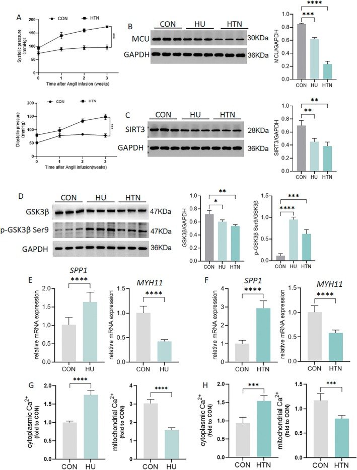
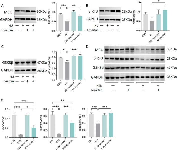
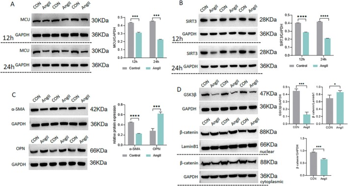
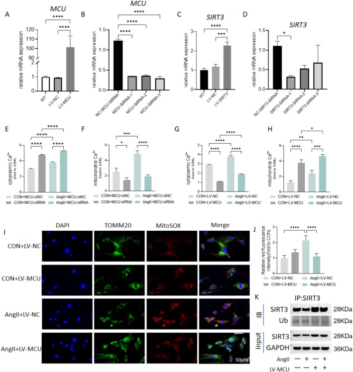
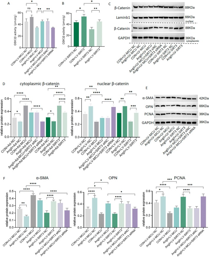
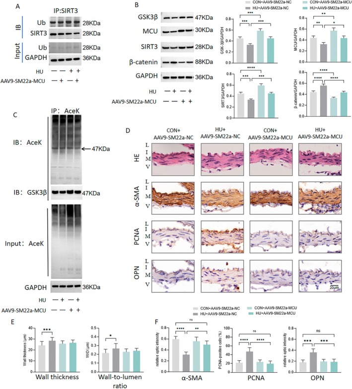
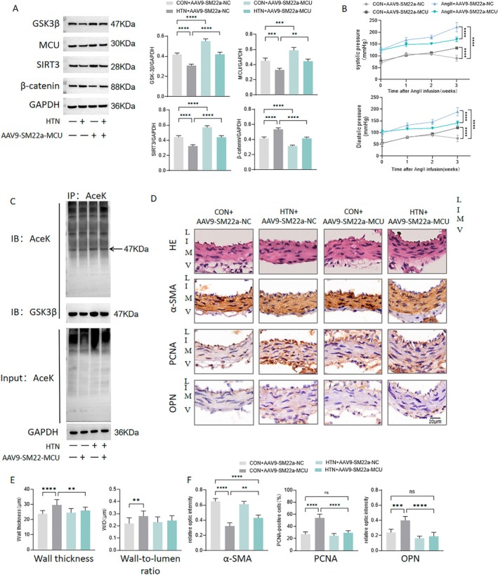
Calcium homeostasis plays a crucial role in regulating the phenotype of vascular smooth muscle cells (VSMCs) and vascular remodeling. This study aims to investigate the role of the mitochondrial calcium uniporter (MCU), which facilitates the uptake of Ca2+ into the mitochondria, in vascular remodeling and its underlying regulatory mechanisms. Vascular remodeling in rats was induced through either 21-day hindlimb unloading (HU) or 21-day angiotensin II (Ang II) infusion (0.7 mg/kg/day). Phenotypic switching of VSMCs and vascular remodeling were assessed. To induce phenotypic switching and clarify the underlying regulatory mechanisms, VSMCs were treated with Ang II (100 μmol/L). Gene manipulation was performed using plasmids, lentivirus, and adeno-associated virus serotype 9 (AAV9). Mitochondrial oxidative stress, Ca2+ distribution, and the expression of MCU, SIRT3, GSK3β, and β-catenin, along with GSK3β activity, SIRT3 ubiquitination, and GSK3β acetylation, were evaluated. The expression of MCU and SIRT3 in rat cerebral arteries was downregulated following HU and Ang II administration, which resulted in an increase in cytoplasmic Ca2+, a decrease in mitochondrial Ca2+, and a shift toward a synthetic phenotype in VSMCs. In vitro, Ang II treatment of VSMCs led to reduced expression of MCU, SIRT3, and GSK3β, and increased nuclear translocation of β-catenin. Knockdown of MCU caused an increase in cytoplasmic Ca2+ and a reduction in mitochondrial Ca2+, while MCU overexpression had the opposite effect, decreasing cytoplasmic Ca2+ and increasing mitochondrial Ca2+. Additionally, MCU overexpression decreased SIRT3 ubiquitination, mitochondrial oxidative stress, GSK3β acetylation, nuclear translocation of β-catenin, and VSMC phenotypic switching-these effects were blocked by SIRT3 knockdown. Moreover, MCU overexpression partially mitigated vascular remodeling in HU and hypertensive rats by inhibiting the GSK3β/β-catenin pathway and preserving SIRT3. Ang II regulates MCU protein expression, which is reduced in the HU and Ang II-induced hypertensive rat cerebral arteries. This reduction impairs cellular Ca2+ buffering and promotes mitochondrial oxidative stress. The stress response triggers the downstream degradation of SIRT3, which subsequently inhibits the activity of GSK3β via acetylation and promotes the nuclear translocation of β-catenin, thereby facilitating phenotypic switching and vascular remodeling.

Keywords: GSK3β/β‐catenin; SIRT3; angiotensin II; calcium; hindlimb unloading; microgravity; mitochondrial calcium uniporter; vascular remodeling.


Conflict of interest statement

The authors declare no conflicts of interest.

Figures

FIGURE 1
FIGURE 1
Expression of MCU, SIRT3, and GSK3β, calcium homeostasis and phenotypic markers in cerebral arteries of rats with hindlimb unloading (HU) and angiotensin II (Ang II)‐induced hypertension. Rats were subjected to 21‐day HU to simulate microgravity effects and treated with a 21‐day Ang II infusion (0.7 mg/kg/day) to establish hypertension. (A) Noninvasive tail cuff monitoring of systolic blood pressure (SBP) and diastolic blood pressure (DBP) in rats with vehicle or Ang II for 21 days. (B–D) Western blot analyses of MCU, SIRT3, GSK3β and p‐GSK3β Ser9 in HU and hypertensive (HTN) rats. (E, F) Quantitative PCR analyses of SPP1 and MYH11 in the cerebral arteries of HU and HTN rats. (G, H) Cytoplasmic and mitochondrial Ca2+ levels in cerebral VSMCs of HU and HTN rats. n = 6. *p < 0.05, **p < 0.01, ***p < 0.001, ****p < 0.0001.
FIGURE 2
FIGURE 2
Expression of MCU, SIRT3, and GSK3β in the cerebral arteries of rats with hindlimb unloading (HU) and angiotensin II (Ang II)‐induced hypertension following Losartan administration. HU and hypertensive rats received distilled water containing losartan at 30 mg/kg/day or vehicle by gavage. (A, B) Western blot analyses of MCU and SIRT3 in HU rats, with or without Losartan administration. (C) Western blot analyses of MCU, SIRT3, and GSK3β in hypertensive rats, with or without Losartan administration. n = 6. *p < 0.05, **p < 0.01, ***p < 0.001, ****p < 0.0001.
FIGURE 3
FIGURE 3
Expression of MCU, SIRT3, α‐SMA, OPN, GSK3β and β‐catenin in vascular smooth muscle cells (VSMCs) incubated In vitro with angiotensin II (Ang II). The VSMCs were incubated In vitro with Ang II (100 nmol/L). (A, B) Western blot analyses of MCU and SIRT3 in Ang II‐treated VSMCs after 12 and 24 h of incubation. (C) Western blot analyses of the contractile marker α‐SMA and synthetic marker OPN in Ang II‐treated VSMCs after 24 h of incubation. (D) Western blot analyses of nuclear and cytoplasmic β‐catenin and GSK3β in Ang II‐treated VSMCs after 24 h of incubation. n = 5. *p < 0.05, ***p < 0.001, ****p < 0.0001.
FIGURE 4
FIGURE 4
Disruption of Ca2+ buffering, mitochondrial oxidative stress, and SIRT3 ubiquitin‐mediated degradation following MCU inhibition in vascular smooth muscle cells (VSMCs). The VSMCs were incubated In vitro with angiotensin II (Ang II) (100 nmol/L). (A–D) Verification of overexpression lentivirus and knockdown plasmids targeting MCU and SIRT3. (E–H) Cytoplasmic and mitochondrial Ca2+ levels in VSMCs treated with Ang II for 24 h, following MCU overexpression and knockdown. (I, J) MitoSOX analysis of mitochondrial ROS in VSMCs treated with Ang II, with or without MCU overexpression. (K) Representative western blot images of VSMCs immunoprecipitated (IP) with SIRT3 and immunoblotted using ubiquitin (Ub) antibodies. Input SIRT3 is also shown. n = 5. *p < 0.05, **p < 0.01, ***p < 0.001, ****p < 0.0001.
FIGURE 5
FIGURE 5
MCU regulates GSK3β acetylation via SIRT3 in vascular smooth muscle cells (VSMCs). The VSMCs were incubated In vitro with angiotensin II (Ang II) (100 nmol/L). (A) Representative western blot images of VSMCs immunoprecipitated with a GSK3β antibody and immunoblotted with GSK3β and SIRT3 antibodies. (B) Representative western blot images of VSMCs immunoprecipitated with a SIRT3 antibody and immunoblotted with GSK3β and SIRT3 antibodies. (C) Western blot analysis of MCU in VSMCs. (D) Western blot analysis of SIRT3 in VSMCs. (E) Representative western blot images of VSMCs immunoprecipitated with a GSK3β antibody and immunoblotted with anti‐acetyl‐lysine (Ace‐K) and GSK3β antibodies. n = 5. *p < 0.05, **p < 0.01, ***p < 0.001, ****p < 0.0001.
FIGURE 6
FIGURE 6
MCU regulates enzymatic activity of GSK3β and phenotype switching of vascular smooth muscle cells (VSMCs) via GSK3β/β‐catenin pathway. The VSMCs were incubated In vitro with angiotensin II (Ang II) (100 nmol/L). (A, B) Enzymatic activity of GSK3β in VSMCs. (C, D) Western blot analysis of nuclear and cytoplasmic β‐catenin in VSMCs. (E, F) Western blot analysis of α‐SMA, OPN and PCNA in VSMCs. n = 5. *p < 0.05, **p < 0.01, ***p < 0.001, ****p < 0.0001.
FIGURE 7
FIGURE 7
MCU regulates cerebrovascular remodeling in hindlimb unloading (HU) rats. Rats were subjected to 21‐day HU to simulate microgravity effects. (A) Representative Western blot images of cerebral arteries immunoprecipitated with a SIRT3 antibody and immunoblotted with ubiquitin (Ub) antibodies. (B) Western blot analysis of MCU, SIRT3, GSK3β, and β‐catenin in HU rat cerebral arteries. (C) Representative western blot images of cerebral arteries immunoprecipitated with a GSK3β antibody and immunoblotted with anti‐acetyl‐lysine (Ace‐K) antibodies. (D) Representative images of H&E staining and representative images of immunohistochemical staining for α‐SMA, OPN, and PCNA of basilar arteries in HU rats. (E) Quantitative analysis of wall thickness and wall‐to‐lumen ratio basilar arteries in HU rats. (F) Quantitative analysis of relative optical density of α‐SMA, OPN, and PCNA, calculated by normalizing the integrated optical density to vessel wall area. L, lumen; I, intima; M, media; and V, adventitia. n = 5. *p < 0.05, **p < 0.01, ***p < 0.001, ****p < 0.0001.
FIGURE 8
FIGURE 8
MCU regulates cerebrovascular remodeling in angiotensin II (Ang II)‐induced hypertensive rats (HTN). Rats were treated with a 21‐day Ang II infusion (0.7 mg/kg/day) to establish hypertension. (A) Noninvasive tail cuff monitoring of systolic and diastolic blood pressure in rats infused with Ang II. (B) Western blot analysis of MCU, SIRT3, GSK3β, and β‐catenin in cerebral arteries of hypertensive rats. (C) Representative Western blot images of cerebral arteries immunoprecipitated with a GSK3β antibody and immunoblotted with anti‐acetyl‐lysine (Ace‐K) antibodies. (D) Representative images of H&E staining and representative images of immunohistochemical staining for α‐SMA, OPN, and PCNA of basilar arteries in hypertensive rats. (E) Quantitative analysis of wall thickness and wall‐to‐lumen ratio basilar arteries in hypertensive rats. (F) Quantitative analysis of relative optical density of α‐SMA, OPN, and PCNA, calculated by normalizing the integrated optical density to vessel wall area. L, lumen; I, intima; M, media; and V, adventitia. n = 5. **p < 0.01, ***p < 0.001, ****p < 0.0001.

Similar articles

References

    1. Scott J. M., Stoudemire J., Dolan L., and Downs M., “Leveraging Spaceflight to Advance Cardiovascular Research on Earth,” Circulation Research 130 (2022): 942–957. - PMC - PubMed
    1. Zhang L., “Region‐Specific Vascular Remodeling and Its Prevention by Artificial Gravity in Weightless Environment,” European Journal of Applied Physiology 113 (2013): 2873–2895. - PubMed
    1. Xie M., Zhang L., Ma J., and Cheng H., “Functional Alterations in Cerebrovascular K+ and Ca2+ Channels Are Comparable Between Simulated Microgravity Rat and SHR,” American Journal of Physiology. Heart and Circulatory Physiology 289 (2005): H1265–H1276. - PubMed
    1. Zhang R., Bai Y. G., Lin L. J., et al., “Blockade of AT1 Receptor Partially Restores Vasoreactivity, NOS Expression, and Superoxide Levels in Cerebral and Carotid Arteries of Hindlimb Unweighting Rats,” Journal of Applied Physiology 106 (2009): 251–258. - PubMed
    1. Zhang R., Jiang M., Zhang J., et al., “Regulation of the Cerebrovascular Smooth Muscle Cell Phenotype by Mitochondrial Oxidative Injury and Endoplasmic Reticulum Stress in Simulated Microgravity Rats via the PERK‐eIF2α‐ATF4‐CHOP Pathway,” Biochimica et Biophysica Acta 1866 (2020): 165799. - PubMed

MeSH terms

LinkOut - more resources