. 2025 Jul 1;28(4):606-608.
doi: 10.4103/aian.aian_747_24. Epub 2025 Jul 7.

Cerebral Chromoblastomycosis: A Unique Presentation of Dematiaceous Fungal Infection in an Immunocompromised Patient

Affiliations

Cerebral Chromoblastomycosis: A Unique Presentation of Dematiaceous Fungal Infection in an Immunocompromised Patient

Norton Stephen et al. Ann Indian Acad Neurol. .
No abstract available


Conflict of interest statement

There are no conflicts of interest.

Figures

Figure 1
Figure 1
CECT (a) and serial slices of cMRI axial view T1 post-contrast images of the brain (b-d) show multiple ring-enhancing lesions in the right parietal, callosal, and pericallosal regions. CECT: Contrast-enhanced computed tomography, cMRI: Contrast magnetic resonance imaging
Figure 2
Figure 2
(a and b) The section shows well-formed epithelioid granulomas surrounded by lymphocytes, along with golden-brown pigmented, thick-walled fungal structures called Medlar bodies, as highlighted by black arrows (H and E; a- 200×, b- 400×). (c) Reactive gliosis in the brain parenchyma (H and E, 400×). (d and e) Sclerotic bodies highlighted by PAS stain and GMS stain (400×). (f) Sclerotic bodies highlighted by Masson-Fontana stain (200×). GMS: Gömöri’s methenamine silver, H and E: hematoxylin and eosin, PAS: periodic acid Schiff
Figure 3
Figure 3
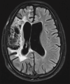
Image of follow-up MRI (FLAIR) after 1 year revealed encephalomalacia with gliotic changes in the right cerebral hemisphere and resolution of the previously observed ring-enhancing lesions. FLAIR: fluid-attenuated inversion recovery, MRI: magnetic resonance imaging

References

    1. Queiroz-Telles F. Chromoblastomycosis: A neglected tropical disease. Rev Inst Med Trop São Paulo. 2015;57:46–50. - PMC - PubMed
    1. Esterre P, Andriantsimahavandy A, Ramarcel ER, Pecarrere JL. Forty years of chromoblastomycosis in Madagascar: A review. Am J Trop Med Hyg. 1996;55:45–7. - PubMed
    1. Queiroz-Telles F, de Hoog S, Santos DW, Salgado CG, Vicente VA, Bonifaz A, et al. Chromoblastomycosis. Clin Microbiol Rev. 2017;30:233–76. - PMC - PubMed
    1. Revankar SG, Sutton DA, Rinaldi MG. Primary central nervous system phaeohyphomycosis: A review of 101 cases. Clin Infect Dis. 2004;38:206–16. - PubMed
    1. Jinkala SR, Basu D, Neelaiah S, Stephen N, Hanuman SB, Singh R. Subcutaneous phaeohyphomycosis: A clinical mimic of skin and soft tissue neoplasms—A descriptive study from India. World J Surg. 2018;42:3861–6. - PubMed