

Endothelial sensitivity to pro-fibrotic signals links systemic exposure to pulmonary fibrosis
- PMID: 40623988
- PMCID: PMC12234957
- DOI: 10.1038/s41419-025-07824-5
Endothelial sensitivity to pro-fibrotic signals links systemic exposure to pulmonary fibrosis
Abstract
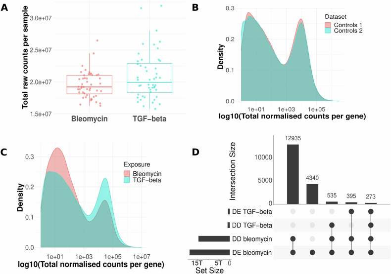
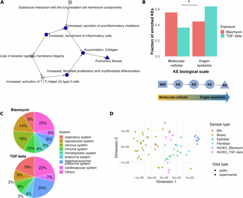
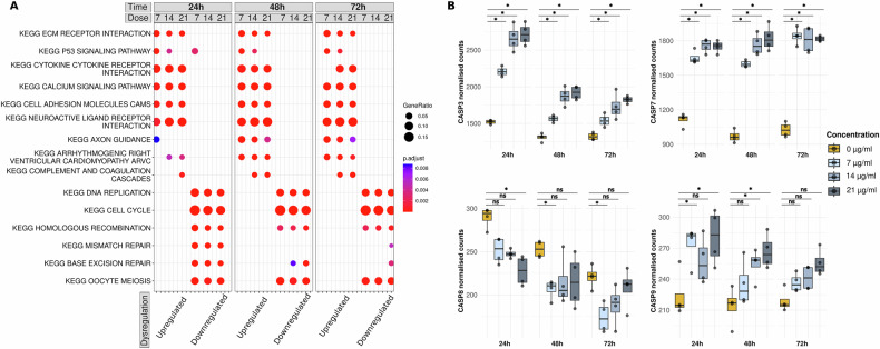
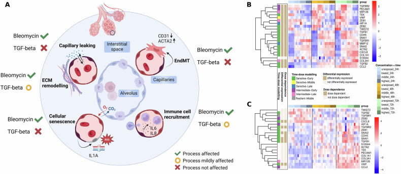
Pulmonary fibrosis (PF) is a life-threatening condition characterised by excessive extracellular matrix deposition and tissue scarring. While much of PF research has focused on alveolar epithelial cells and fibroblasts, endothelial cells have emerged as active contributors to the disease initiation, especially in the context of systemic exposure to pro-fibrotic substances. Here, we investigate early transcriptomic and secretory responses of human umbilical vein endothelial cells (HUVEC) to subtoxic doses of bleomycin, a known pro-fibrotic agent, and TGF-beta, a key cytokine in fibrosis. Bleomycin exposure induced a rapid and extensive shift in the endothelial transcriptional programme, including signatures of endothelial to mesenchymal transition, cellular senescence, and immune cell recruitment. These findings suggest endothelial cells as early initiators of pro-fibrotic signals, independent of contributions from other cell types. In contrast, TGF-beta effects were limited and transient, indicating its pro-fibrotic action may require another initial stimulus and interplay with other cells like fibroblasts. This study highlights the sensitivity of endothelial cells to pro-fibrotic exposure and provides a blueprint of early pro-fibrotic mechanisms that may operate on organs such as the lungs systemically via the endothelium, emphasising its pivotal role in PF pathogenesis.
© 2025. The Author(s).
Conflict of interest statement
Competing interests: The authors declare no competing interests. Ethics approval and consent to participate: The authors confirm that all methods were performed in accordance with the relevant guidelines and regulations. The study exclusively used in vitro models, with no human or animal subjects involved. Declaration of generative AI and AI-assisted technologies in the writing process: During the preparation of this work the authors used ChatGPT to improve the readability of single sentences and to shorten single paragraphs with limited word count allowed. After using this tool, the authors reviewed and edited the content as needed and take full responsibility for the content of the published article.
Figures
References
-
- Dymacek JM, Snyder-Talkington BN, Raese R, Dong C, Singh S, Porter DW, et al. Similar and differential canonical pathways and biological processes associated with multiwalled carbon nanotube and asbestos-induced pulmonary fibrosis: a 1-year postexposure study. Int J Toxicol. 2018;37:276–84. - PMC - PubMed
-
- Bhattacharya M, Ramachandran P. Immunology of human fibrosis. Nat Immunol. 2023;24:1423–33. - PubMed
MeSH terms
Substances
LinkOut - more resources
Full Text Sources
Medical
