. 2025 Jul 8;16(1):6271.
doi: 10.1038/s41467-025-61587-3.

Rab27a regulates the transport of influenza virus membrane proteins to the plasma membrane

Affiliations

Rab27a regulates the transport of influenza virus membrane proteins to the plasma membrane

Tong Chen et al. Nat Commun. .

Abstract

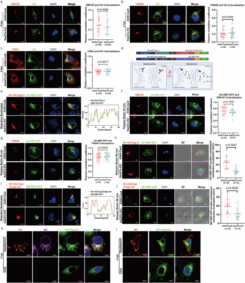
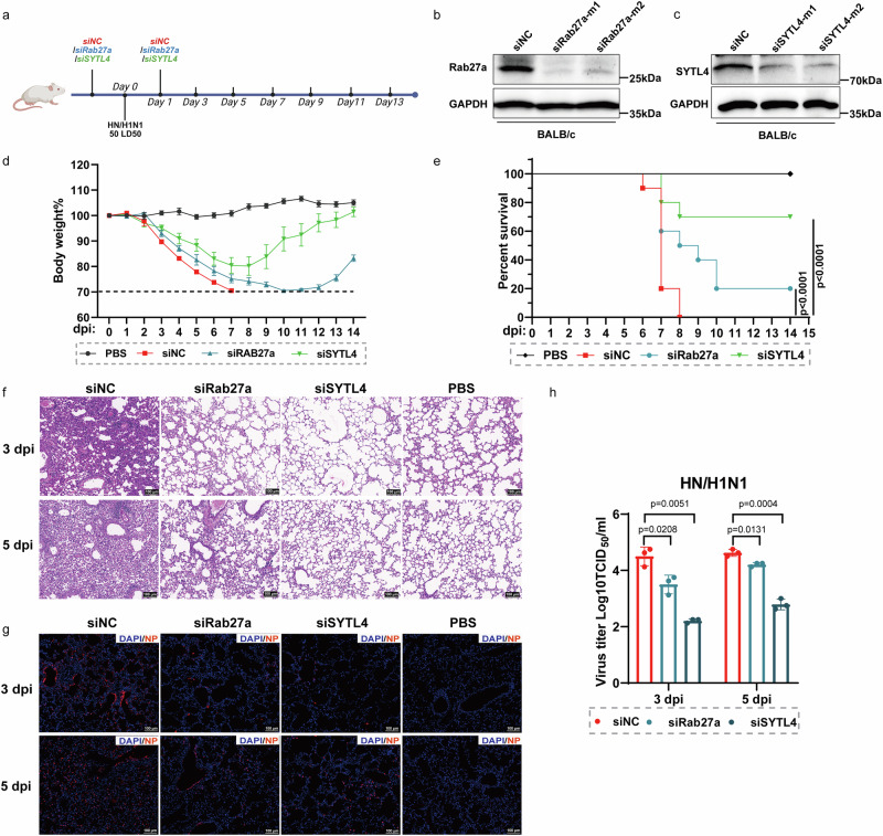
The molecular mechanisms underlying the transport of influenza A virus (IAV) membrane proteins to the cell surface remain largely unclear. In this study, siRNA screening identifies Rab27a as a critical host factor regulating this transport process. GTP-bound Rab27a operates via its effectors, synaptotagmin-like protein 1 (SYTL1) and SYTL4, to facilitate the transport of vesicles carrying viral membrane proteins to the plasma membrane. Absence of Rab27a or SYTL4 does not block the early stages of the IAV life cycle but restricts viral assembly and budding. Notably, silencing SYTL4 provides superior protection in the female mouse IAV infection model. This investigation elucidates the molecular mechanism by which Rab27a and its effectors modulate the transport of IAV membrane proteins, thereby bridging a critical gap in IAV life cycle research and presenting a potential target for the development of antiviral drugs.


Conflict of interest statement

Competing interests: The authors declare no competing interests.

Figures

Fig. 1
Fig. 1. Rab27a promotes IAV replication.
a A549 cells were transfected with siNC or siRNAs targeting Rab3b (siRab3b-1, siRab3b-2), Rab6a (siRab6a-1, siRab6a-2), Rab11a (siRab11a-1, siRab11a-2), Rab17 (siRab17-1, siRab17-2), Rab23a (siRab23a-1, siRab23a-2), Rab25 (siRab25-1, siRab25-2), Rab27a (siRab27a-1, siRab27a-2), Rab37 (siRab37-1, siRab37-2), or Rab38 (siRab38-1, siRab38-2) for 24 h. The cells were then infected with HM virus (MOI, 10), and HA protein levels on the cell surface were quantified by flow cytometry at 4 hpi. b A549 cells transfected with siNC or siRab27a (siRab27a-1, siRab27a-2) for 24 h were infected with HM virus (MOI, 0.1). c–e Rab27a KO and A549-Cas9 cells were infected with HM virus (MOI, 0.1), SH13/H9N2 virus (MOI, 0.01), or PR8/H1N1 virus (MOI, 0.01). f–h A549 cells were transfected with 2 μg/ml exogenous Rab27a or an empty vector as a negative control for 24 h. The cells were then infected with HM virus (MOI, 0.1), SH13 virus (MOI, 0.01), or PR8 virus (MOI, 0.01). i A549-Cas9, Rab27a KO, and Rab27a KO cells stably expressing Flag-Rab27a-WT, Flag-Rab27a-T23N (dominant-negative mutant), or Flag-Rab27a-Q78L (constitutively active mutant) were infected with HM virus (MOI, 0.1). Rab27a and NP protein expression levels were examined by western blot (b, f, g, h, and i), with GAPDH or β-actin serving as a loading control. Viral titers in the supernatants were quantified using a TCID50 assay on MDCK cells (b–i). Data represent the means ± SD from three independent experiments. Statistical significance was determined using a two-tailed Student’s t-test.
Fig. 2
Fig. 2. Knocking out Rab27a restricts IAV assembly and budding.
a Rab27a-KO and A549-Cas9 cells were infected with HM virus (MOI, 10). At 0.5 and 1 hpi, cells were fixed, permeabilized, and stained with anti-NP (red) and DAPI (blue). At least 9 randomly selected fields from each group were analyzed for quantitative co-localization of the nucleus and NP protein. Representative images are shown in (a). Scale bar, 20  μm. Data represent the mean ± SD from one representative experiment of at least three independent experiments, with at least 100 cells per condition quantified. b–e Rab27a-KO and A549-Cas9 cells were pretreated with cycloheximide (CHX, 100 μM) or DMSO (−CHX) for 4 h, followed by infection with HM virus (MOI, 10) and subsequent treatment with CHX (350 μM) or DMSO. The mRNA and vRNA levels of viral genes were quantified by qRT-PCR and normalized to 18S rRNA levels. f A549-Cas9, Rab27a-KO, and Rab27a-KO cells stably expressing Flag-Rab27a-WT, Flag-Rab27a-T23N, or Flag-Rab27a-Q78L were infected with HM virus (MOI, 10). Cells were fixed in glutaraldehyde at 6 hpi, and the budding of influenza viruses was observed using transmission electron microscopy (TEM). For quantitative analysis, 30 cells were randomly selected from each group to determine the number of IAV budding events. Representative images are shown in (f), with scale bars of 5 μm and 1 μm. Data represent the mean ± SD from one representative experiment of at least three independent experiments. Statistical significance was analyzed using a two-tailed Student’s t-test.
Fig. 3
Fig. 3. Rab27a regulates transport of HA and NA proteins to apical plasma membrane.
a Rab27a-KO and A549-Cas9 cells were infected with HM virus (MOI, 10). Rab27a and viral protein levels were analyzed by western blot at 0, 3, 6, and 9 hpi, with GAPDH serving as a loading control. b Western blot densitometry at 6 hpi analysis was performed using ImageJ software. c–h Rab27a-KO and A549-Cas9 cells were infected with HM virus (MOI, 10). At 6 hpi, total (permeabilized) and cell surface (nonpermeabilized) levels of viral membrane proteins were quantified by flow cytometry. The bar graph shows the MFI. i–k Rab27a-KO and A549-Cas9 cells were infected with HM virus (MOI, 10). At 6 hpi, cells were fixed, permeabilized, and stained with antibodies against HA, NA, and M2 (all in red) and DAPI (blue). Scale bar, 10 μm. l, m A549-Cas9, Rab27a-KO, and Rab27a-KO cells stably expressing Flag-Rab27a-WT, Flag-Rab27a-T23N, or Flag-Rab27a-Q78L were infected with HM virus (MOI, 10). At 6 hpi, HA protein levels on the cell surface were quantified by flow cytometry (l), while HA, NP, and Rab27a protein levels were analyzed by western blot (m), with β-actin serving as a loading control. n, o A549 cells transfected with siNC or siRab27a (siRab27a-1, siRab27a-2) for 24 h were infected with VSV (MOI, 10). Total (n) and cell surface (o) levels of VSV-G protein were quantified by flow cytometry. Data are presented as mean ± SD from three independent experiments. Statistical significance was determined using a two-tailed Student’s t-test.
Fig. 4
Fig. 4. Rab27a promotes autophagy and M2 transport to plasma membrane.
a HEK293T cells were transfected with Rab27a or vector control for 24 h, infected with HM virus (MOI, 0.1) or left uninfected, and treated with BafA1 (400 nM) or DMSO for 24 h. b Rab27a-KO and A549-Cas9 cells were infected with HM virus (MOI, 0.1) or left uninfected and treated with BafA1 (400 nM) or DMSO for 24 h. c Rab27a-KO and A549-Cas9 cells were transfected with GFP-LC3 for 24 h, followed by starvation treatment for another 24 h. Scale bar, 10 μm. d Rab27a-KO and A549-Cas9 cells were infected with HM virus (MOI, 0.1) or left uninfected for 6 and 12 hpi. e Molecular modeling predicts complex formation involving ATG4B (golden), Rab27a (orange), and LC3 (green) proteins, highlighting the phosphorylation site serine 316 (Ser316, red) in ATG4B. f, g HEK293T cells were co-transfected with Rab27a, ATG4B or empty vector for 36 h. co-IP was performed using an anti-Flag antibody (f). Cells were treated with CQ (100 μM) (g). h Rab27a-KO and A549-Cas9 cells were infected with HM virus (MOI, 0.1) or left uninfected for 12 hpi. i HEK293T cells were co-transfected with ATG4B and Rab27a or empty vectors. At 24 h post-transfection, cells were subjected to 12 h of starvation. j HEK293T cells were co-transfected with ATG4B and Rab27a along with M2 (0, 0.5, or 1 μg/ml) or empty vectors. At 24 h post-transfection, cells were subjected to 12 h of starvation. k A549 cells were co-transfected with HA-Rab27a and LC3-GFP for 36 h, pretreated with CHX (100 μM) or DMSO for 4 h, and then infected with HM virus (MOI, 10). Cells were further treated with CHX (350 μM) or DMSO and fixed at 3 hpi. Staining with anti-HA, anti-M2, and DAPI. Scale bar, 10 μm. l HEK293T cells were co-transfected with Rab27a, M2 for 24 h. Total and cell surface levels of M2 protein were quantified by flow cytometry. Data are presented as mean ± SD from three independent experiments. Statistical significance was assessed using a two-tailed Student’s t-test. Western blot analysis was performed for the indicated proteins, with GAPDH or β-actin as a loading control.
Fig. 5
Fig. 5. Rab27a regulates the transport of viral membrane proteins to the plasma membrane through Rab27a positive vesicles.
a–c Rab27a-KO and A549-Cas9 cells were infected with HM virus (MOI, 10). At 3 (c) and 4 hpi (a, b), cells were stained with anti-GM130, TGN46, CD63, and influenza HA. The co-localization of HA with different organelles was quantified by calculating the Pearson correlation coefficient (PCC) using the Coloc 2 in Fiji. GM130 (a, cis-Golgi, n = 20); TGN46 (b, trans-Golgi, n = 25); CD63 (c, endosome, n = 22). d i. Schematic of genes coding for the hook (Str-HA (Tag) -Li/Golgin-84) and the reporter (HA/M2/VSV G-SBP-GFP), expressed under the same CMV promoter, separated by an internal ribosome entry site (IRES); ii. Schematic of the RUSH system. In the absence of biotin, SBP-tagged cargo is retained in the ER or Golgi via interaction with streptavidin-HA (Tag) -Li (ER-localized)/Golgin-84 (Golgi-localized). Addition of biotin disrupts the interaction, allowing forward trafficking of cargo. Image was created in BioRender. Tong, C. (2025) https://BioRender.com/nuf5a1w. e–j cells were transfected with Str-HA (Tag) -Li_HA-SBP-GFP or Str-HA (Tag) -Golgin-84_M2-SBP-GFP for 24 h, followed by addition of biotin (400 μM). At corresponding time after biotin addition, cells were stained with anti-HA (Tag), GM130, and TGN46. Co-localizations of M2/HA with ER (e, i) or proportion of HA/M2 on the cell surface (h, j, n = 15,) was analyzed using Fiji. The co-localization of HA-SBP-GFP with different organelles was also quantified by calculating the PCC. GM130 (f, cis-Golgi, n = 21); TGN46 (g, trans-Golgi, n = 28). k, l A549 cells stably expressing GFP-Rab27a were pretreated with CHX (100 μM) or DMSO for 4 h, and then infected with HM virus (MOI, 10). Cells were further treated with CHX (350 μM) or Oseltamivir (120 μM) and fixed at 4 hpi. In (k), staining with anti-HA, anti-NA, and DAPI. In (l), staining with anti-M2 and DAPI. White arrows indicate colocalization of HA, NA, and GFP-Rab27a; blue arrows indicate colocalization of NA and GFP-Rab27a; yellow arrows indicate colocalization of HA and GFP-Rab27a in (k), and of M2 and GFP-Rab27a in (l). Scale bar, 10 μm. Representative images are shown in (a–c, e–l). Data are presented as mean ± SD from three independent experiments. Statistical significance was assessed using a two-tailed Student’s t-test.
Fig. 6
Fig. 6. SYTL1 and SYTL4 are two Rab27 effectors involved in IAV membrane protein transport.
a HEK293T cells were transfected with Rab27a-GFP for 24 h, then infected with HM virus (MOI, 0.1) or left uninfected. At 12 hpi, cell lysates were subjected to GST affinity pull-down assays. b HEK293T cells were transfected with Flag-Rab27a WT, Flag-Rab27a-Q78L, and Flag-Rab27a-T23N for 24 h, then cell lysates were used for GST affinity pull-down assays. c, d A549 cells transfected with siSYTL1/4 or siNC for 24 h were infected with HM virus. Protein levels were detected at 6 hpi (c) (MOI, 10), and viral titers of supernatants were determined by TCID50 assay at 24 hpi (d) (MOI, 0.1). e, f SYTL1/4-KO and A549-Cas9 cells were infected with HM virus. Protein levels were detected at 6 hpi (e) (MOI, 10), and viral titers of supernatants were determined by TCID50 assay at 18 and 36 hpi (f) (MOI, 0.1). gk SYTL4-KO and A549-Cas9 cells were pretreated with CHX (100 μM) or DMSO for 4 h, then infected with HM virus (MOI, 10) and treated with CHX (350 μM) or DMSO. mRNA and vRNA levels of viral genes were detected by qRT-PCR. l SYTL4-KO and A549-Cas9 were pretreated with CQ (12.5 μM) or DMSO for 6 h, then cells infected with HM virus (MOI, 10) and treated with CQ (25 μM) for 6 h. m–p SYTL4-KO and A549-Cas9 cells were pretreated with CQ (12.5 μM) or DMSO for 6 h, then infected with HM virus (MOI, 10) and treated with CQ (25 μM). At 6 hpi, both total (m, o) and cell surface (n, p) viral membrane proteins levels were measured by flow cytometry. q, r A549 cells transfected with siSYTL4-1, siSYTL4-2, or siNC for 24 h were infected with VSV-GFP (MOI, 10). Cell surface (q) and total (r) levels of VSV-G protein were quantified by flow cytometry. Data are presented as mean ± SD from three independent experiments. Statistical significance was determined using a two-tailed Student’s t-test. Levels of indicated proteins, assessed by western blot in (ac, e, and l), GAPDH served as a loading control.
Fig. 7
Fig. 7. SYTL4 is a potential anti-influenza virus drug target.
a A flowchart of siRNA treatment and HN/H1N1 infection in a mouse experimental model. Image was created in BioRender. Tong, C. (2025) https://BioRender.com/hq3p0b8. b, c Western blot analysis of Rab27a expression in siRab27a (b), siSYTL4 (c) and siNC treated mice at 3 dpi, GAPDH served as a loading control. d Body weight changes of PBS and HN/H1N1 infection with siRNAs treatment in mice (n = 10). Mice with body weight below 70% of initial were euthanized according to the ethical principles of animal welfare. e Survival rates of PBS and HN/H1N1-infected mice with siRNAs treatment (n = 10). Survival curves were analyzed using the log-rank test (Mantel-Cox). f Pathological lesions in the lungs of mice infected with the indicated virus at 3 and 5 dpi with hematoxylin and eosin (H&E) staining. Scale bars, 100 μm. g Immunofluorescent staining of lung sections from mice infected with the indicated virus at 3 and 5 dpi. Viral NP antigen was stained red, and the cell nucleus was stained blue. Scale bars, 100 μm. h Virus titers in the lungs of infected mice (n = 3) at 3 and 5 dpi. The data shown in (d and h) are means ± SD (n  =  10 for (c) and n  =  3 for (h) biologically independent experiments). Statistical significance was analyzed using a two-tailed Student’s t-test.

References

    1. Iuliano, A. D. et al. Estimates of global seasonal influenza-associated respiratory mortality: a modelling study. Lancet391, 1285–1300 (2018). - PMC - PubMed
    1. Rossman, J. S. & Lamb, R. A. Influenza virus assembly and budding. Virology411, 229–236 (2011). - PMC - PubMed
    1. Eisfeld, A. J., Kawakami, E., Watanabe, T., Neumann, G. & Kawaoka, Y. RAB11A Is Essential for transport of the influenza virus genome to the plasma membrane. J. Virol.85, 6117–6126 (2011). - PMC - PubMed
    1. Amorim, M. J. et al. A Rab11-and microtubule-dependent mechanism for cytoplasmic transport of influenza A virus viral RNA. J. Virol.85, 4143–4156 (2011). - PMC - PubMed
    1. Avilov, S. V., Moisy, D., Naffakh, N. & Cusack, S. Influenza A virus progeny vRNP trafficking in live infected cells studied with the virus-encoded fluorescently tagged PB2 protein. Vaccine30, 7411–7417 (2012). - PubMed

MeSH terms

LinkOut - more resources