. 2025 Jul 7;16(1):501.
doi: 10.1038/s41419-025-07803-w.

eEF2K is a poor prognostic factor and novel molecular target in pancreatic cancer: regulating tumor growth and progression via the tumor microenvironment

Affiliations

eEF2K is a poor prognostic factor and novel molecular target in pancreatic cancer: regulating tumor growth and progression via the tumor microenvironment

Didem Karakas et al. Cell Death Dis. .

Abstract

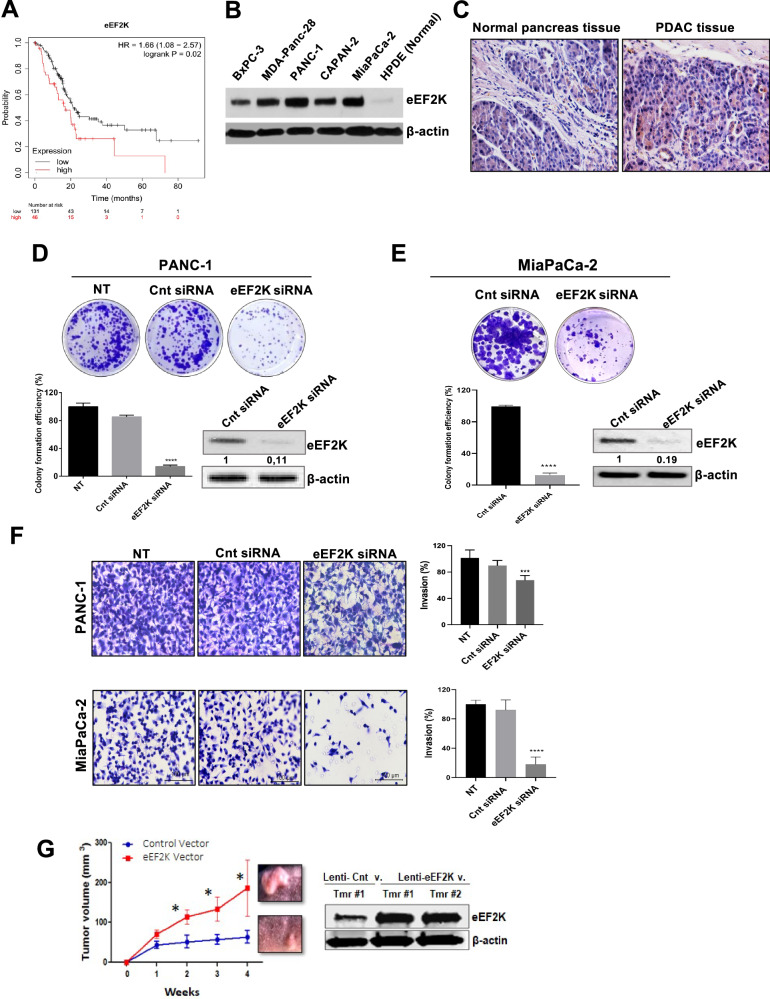
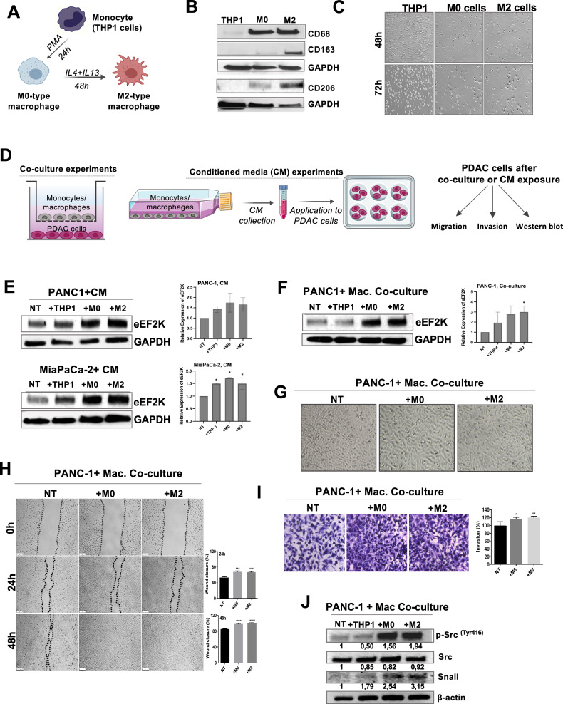
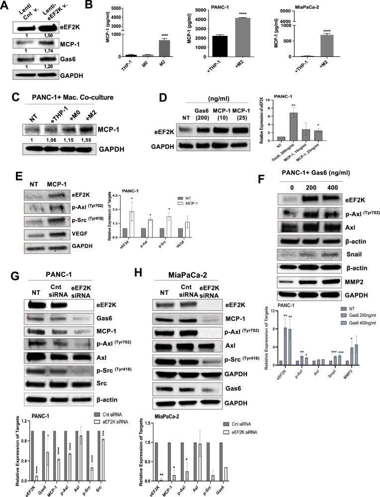
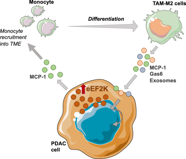
Pancreatic ductal adenocarcinoma (PDAC) is one of the most lethal cancers, with an average survival time of only six months following diagnosis, even with currently available therapies. Thus, PDAC represents a significant therapeutic challenge, necessitating a deeper understanding of its biology and tumor microenvironment (TME) to develop more effective treatments and improve patient outcomes. Here, we report that the expression of Eukaryotic Elongation Factor-2 Kinase (eEF2K) is associated with shorter patient survival and demonstrate that eEF2K signaling is critical for the PDAC tumor growth and regulated by the TME. Furthermore, in vivo targeted genetic inhibition of eEF2K suppressed tumor growth in two different PDAC mouse models, reduced tumor-associated macrophages (TAMs), and induced marked apoptosis in tumor tissues without any signs of toxicity. Our data suggest that eEF2K knockdown diminishes the activity of the AXL receptor tyrosine kinase and reduces the expression of macrophage-derived factors, such as Monocyte Chemoattractant Protein-1 (MCP1), along with the Gas6/AXL signaling pathway in PDAC cells. Additionally, analysis of the NCI-TCGA PDAC patient database further showed that eEF2K expression, in the presence of TAM markers, correlates with even shorter patient survival. TAM-released factors, such as MCP1, Gas6, and exosomes, induce eEF2K expression in PDAC cells, as well as the activity of AXL, SRC, VEGF, Snail, and MMP2, contributing to epithelial-to-mesenchymal transition (EMT), invasion, metastasis, and angiogenesis. In conclusion, our findings reveal for the first time that eEF2K is a critical oncogenic driver of PDAC tumor growth and thus targeting eEF2K represents a promising and novel therapeutic strategy for PDAC.


Conflict of interest statement

Competing interests: The authors declare no competing interests. Ethics approval and consent to participate: Animal experiments were performed in accordance with institutional guidelines and regulations. The animal study protocol was approved by the MD Anderson Cancer Center Animal Research Ethics Committee. Written informed consent was obtained from participants for the publication of identifiable images included in the manuscript.

Figures

Fig. 1
Fig. 1. eEF2K expression is associated with poor overall survival in PDAC patients and aggressive properties of pancreatic cancer cells.
A Higher expression of eEF2K is correlated with poor overall survival (OS) in PDAC patients as determined by Kaplan-Meier survival analysis (P = 0.02). The numbers of patients with low and high eEF2K expression are presented at the bottom of the graph. Mo, months. B eEF2K expression in PDAC cell lines (BxPC-3, MDA-Panc-28, PANC-1, Capan2, MiaPaCa-2) compared to normal pancreatic duct epithelial (HPDE) cells was determined by Western blot analysis. C eEF2K expression in pancreatic cancer patient tissues and normal tissues was determined by immunohistochemistry. D, E eEF2K knockdown by eEF2K siRNA in PANC-1 and MiaPaCa-2 suppressed the colony-forming capability of the PDAC cells. Upper panels show representative images of the colonies and lower panels show quantification of the number of colonies formed (****P < 0.0001, relative to cnt siRNA). The data in the bar graphs represent the mean of three independent experiments. F Knockdown of eEF2K by siRNA inhibits in vitro invasion capability of PDAC cells in Matrigel (***P < 0.001). Bar graphs represent the mean of three independent experiments and seven fields per sample. G Lenti-based ectopic overexpression of human eEF2K gene in PANC-1 cells promoted in vivo tumor xenografts (n = 5 mice/group, *P < 0.05). eEF2K overexpression was confirmed in PDAC tumor xenografts obtained from PANC-1 cell-bearing tumors by Western blot. β-actin was used as a loading control. Cnt: control; v.: vector. Error bars represent ± Standard Deviation (SD).
Fig. 2
Fig. 2. In vivo targeting of eEF2K suppresses tumor growth and decreases MCP-1 expression in orthotopic mouse models of PDAC.
A, B PANC-1 or MiaPaCa-2 tumor-bearing mice were injected (intravenously from the tail vein) with nanoliposomes (NL) incorporating eEF2K siRNA (0.3 mg/kg, n = 5 mice) or control siRNA (0.3 mg/kg, n = 5 mice), once per week, and tumor size was measured weekly by a caliper and mean values presented for each group (*P < 0.05). C The possible toxic effects of eEF2K silencing were investigated by measuring some parameters related to kidney and liver functions (BUN, creatinine, total bilirubin, total protein, alkaline phosphatase, ALT, AST, LDH levels). D Tumor samples from the NL-control siRNA and NL-eEF2K siRNA-treated mice were stained with eEF2K and specific antibodies to detect Ki67, an intratumoral proliferation marker, and CD31, an endothelial marker for evaluation of angiogenesis, and F4/80 for the infiltration of pro-tumorigenic M2 macrophages in PANC-1 tumors. TUNEL staining used for detection of apoptotic cells. Eight randomly selected fields were counted and analyzed for each treatment group (n = 5 mice per group). (Magnification, 20x). TUNEL-positive cells stained (green) in both treatment groups were counted and quantified. Nuclei were stained with DAPI (blue) (**P < 0.01, ***P < 0.001). E Western blot analysis of MCP-1 expression in tumor samples after in vivo targeting of eEF2K. Error bars represent ± SD.
Fig. 3
Fig. 3. Co-culture of PDAC cells with macrophages induces eEF2K expression, migration, and invasion capabilities of cancer cells.
A–C Schematic illustration of monocyte-macrophage differentiation and characterization of macrophage differentiation. B Monocyte-macrophage differentiation was confirmed by Western blot. CD68 was used as a pan-macrophage marker. CD163 and CD206 were used for the confirmation of M2-type macrophage differentiation. C Morphological changes during monocyte-macrophage differentiation were observed under phase-contrast microscope. While monocytes grew in suspension, they became adherent when they polarized into macrophages. M2 pro-tumorigenic macrophages were observed to have a more fibroblast-like phenotype compared to M0 mature macrophages. D Illustration of co-culture and conditioned media experiments to investigate the interactions between PDAC cells and monocytes/macrophages. E The effect of monocyte/macrophage-derived conditioned media (CM) on eEF2K expression of pancreatic cancer cells (PANC-1 and MiaPaCa-2) was evaluated by Western blot (n = 2, *P < 0.05). F The impact of co-culture conditions on eEF2K expression of PANC-1 cells. The changes in eEF2K expression in PANC-1 cells were measured by Western blot after 48 hours of co-culture with monocytes/macrophages (n = 2, *P < 0.05). G The changes in cell morphology after co-culture of PANC-1 cells with macrophages were evaluated through phase-contrast microscopy. H The effect of macrophages on the migration capability of PANC-1 cells was evaluated by wound healing assay (***P < 0.001, ****P < 0.0001, relative to NT). I Cell invasion was evaluated by Matrigel invasion assay in PANC-1 cells (*P < 0.05, **P < 0.01, relative to NT). J Co-culture of macrophages with PANC-1 cells induce signaling pathways and mediators of cell proliferation, survival, cell motility/invasion and epithelial-mesenchymal transition (EMT), including p-Src and Snail detected by Western blot analysis. β-actin and GAPDH were used as loading controls. NT: non-treated; THP-1: monocytes; M0: mature macrophages; M2: tumor-promoting macrophages; CM: conditioned media. Error bars represent ± SD.
Fig. 4
Fig. 4. Macrophage-derived exosomes induce eEF2K expression, cell migration, and invasion in PDAC cells.
A Schematic illustration of exosome experiment design. B Macrophage-derived exosomes induced eEF2K expression, C cell proliferation and colony formation, D cell migration and E invasion of PANC-1 cells compared to non-treated cells (NT) (*P < 0.05, **P < 0.01, ***P < 0.001, ****P < 0.0001, respectively). All experiments were independently confirmed at least two times. F Heat map of unbiased RPPA analysis of PANC-1 cells after exposure to the monocyte or macrophage-derived exosomes. Average log value from fold-change in the expression of indicated proteins identified from a pool of 305 primary antibodies such as 1. MAPK (p-ERK-T202-Y204); 2. Vimentin; 3. YAP (pS127); 4. AMPK (pT172); 5. eIF4G; 6. Myosin-IIa (pS1943); 7. Stat3 (pY705) and 8. B-Raf (pS445). NT: non-treated; THP-1: monocytes; M0: mature macrophages; M2: tumor-promoting macrophages. Error bars represent ± SD.
Fig. 5
Fig. 5. Macrophage-derived factors MCP-1 and Gas6 increase in eEF2K overexpressed cells and these factors, in turn, induce eEF2K expression in PDAC cells.
A Increased MCP-1 and Gas6 protein expressions were detected in eEF2K-overexpressed cells. B Released MCP-1 protein levels in THP-1, M0-type, and M2-type macrophages were measured by ELISA (****P < 0.0001). Besides, MCP-1 levels were measured after co-culturing of PANC-1 or MiaPaCa-2 cells or PSCs with monocytes/macrophages after 48 hours. (****P < 0.0001). C Intracellular MCP-1 levels were measured via Western blot in PANC-1 cells following 48 h co-culture with monocytes or macrophages. D eEF2K protein expression levels were measured by Western blot in PANC-1 cells after Gas6 and MCP-1 exposure (30 minutes and 24 hours, respectively) (n = 2, *P < 0.05, **P < 0.01). E The changes in the expression of eEF2K, p-AxlTyr702, p-SrcTyr416, and VEGF in PANC-1 cells after 24 hours of MCP-1 treatment (n = 2, *P < 0.05). F The effects of different concentrations of Gas6 on the expression of eEF2K, p-AxlTyr702, total Axl, Snail, MMP-2, p-eEF2Thr56 protein levels were evaluated by Western blot (n = 2, *P < 0.05, **P < 0.01, ***P < 0.001). G, H The changes in the expression of eEF2K, Gas6, MCP-1, p-AxlTyr702, total Axl, p-SrcTyr416, total Src, and integrin β1 in PDAC cells following transfection with eEF2K siRNA for 48 h (n = 2, *P < 0.05, **P < 0.01, ***P < 0.001, ****P < 0.0001). Error bars represent ± SD.
Fig. 6
Fig. 6. eEF2K-mediated regulation of the TME.
Schematic representation of a proposed positive feedback loop between tumor cells and TAMs, mediated by the eEF2K/MCP-1 axis. This loop promotes the recruitment and accumulation of monocytes/TAMs, contributing to an immunosuppressive, pro-tumorigenic TME and enhancing pancreatic tumor progression.

Similar articles

References

    1. Siegel R, Naishadham D, Jemal A. Cancer statistics, 2012. CA Cancer J Clin. 2012;62:10–29. - PubMed
    1. Nagendram S, Bhattacharya S. Pancreatic cancer: a glimmer of hope. Trends Urol Men’s Health. 2023;14:5–10.
    1. Garrido-Laguna I, Hidalgo M. Pancreatic cancer: from state-of-the-art treatments to promising novel therapies. Nat Rev Clin Oncol. 2015;12:319–34. - PubMed
    1. Feig C, Gopinathan A, Neesse A, Chan DS, Cook N, Tuveson DA. The Pancreas Cancer Microenvironment. Clin Cancer Res. 2012;18:4266–76. - PMC - PubMed
    1. Delitto D, Black BS, Sorenson HL, Knowlton AE, Thomas RM, Sarosi GA, et al. The inflammatory milieu within the pancreatic cancer microenvironment correlates with clinicopathologic parameters, chemoresistance and survival. BMC Cancer. 2015;15:783. - PMC - PubMed

MeSH terms

Substances