Review
. 2025 Jul 7;30(1):77.
doi: 10.1186/s11658-025-00753-3.

AKT and DUBs: a bidirectional relationship

Affiliations
Review

AKT and DUBs: a bidirectional relationship

Valentina Serratore et al. Cell Mol Biol Lett. .

Abstract

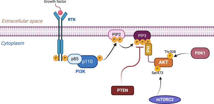
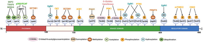
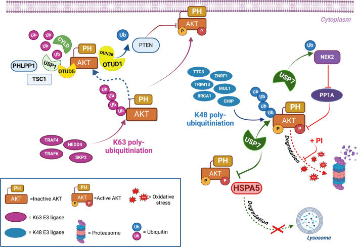
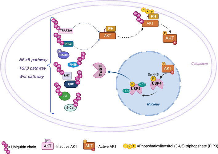
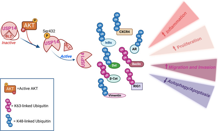
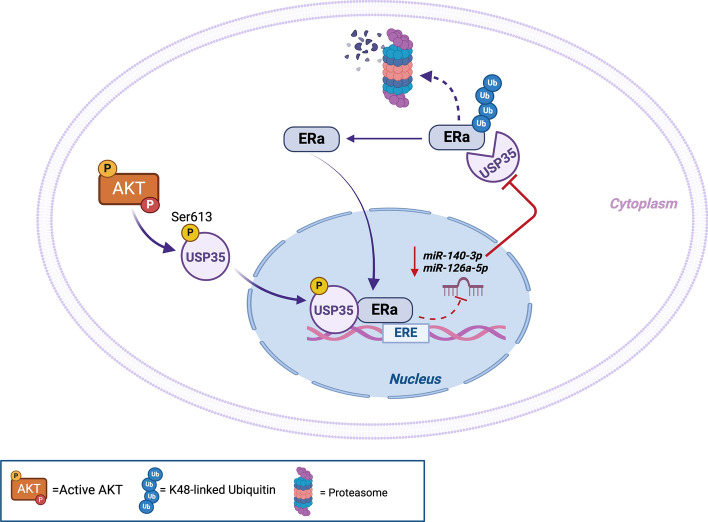
The serine/threonine kinase Akt is crucial for cell physiology and can also contribute to pathology if its activation and regulation is disturbed. This kinase phosphorylates several substrates involved in mechanisms that are altered in human disease. AKT is regulated by several post-translational modifications (PTMs), including ubiquitination/deubiquitination. Ubiquitination can both target AKT to the proteasome and promote its activation. The interplay with the deubiquitination mechanism plays a crucial role in almost all biological activities of AKT. Information on the mechanisms of AKT deubiquitination and its key players has evolved rapidly in recent years along with the development of potential targeting strategies, although many of them are still unclear. Nevertheless, AKT in turn regulates various deubiquitinases (DUBs), suggesting further targeting strategies for human diseases. In this review, we aim to provide an up-to-date overview of the dual relationship between AKT and DUBs with respect to potential translational aim.

Keywords: AKT kinase; Deubiquitinases; Phosphorylation; Post-translational modifications.


Figures

Fig. 1
Fig. 1
The PI3K–AKT pathway. Schematic representation of the PI3K–AKT signaling pathway. Tyrosine kinase receptors (RTK) recruit the regulatory subunit of phosphatidyl inosiltol 3-kinase (PI3K), p85, with subsequent activation of the catalytic subunit, p110. The activated PI3K phosphorylates the (4,5)-biphosphates of phosphatidylinositol (PIP2) to (3,4,5)-triphosphates (PIP3), which in turn recruit AKT via binding to its pleckstrin homology (PH) domain. In this way, AKT could be activated by phosphorylation of Thr308 and Ser473. The phosphatase PTEN is one of the key negative regulators of the pathway as it dephopshorylates PIP3 to PIP2. Created with BioRender.com
Fig. 2
Fig. 2
AKT post-translational modifications (PTMs). The figure shows the PTMs of AKT and their distribution along the different functional domains of the kinase. The residues of the isoform AKT1 are shown. Created with BioRender.com
Fig. 3
Fig. 3
Classification and domain architecture of the DUB families. The deubiquitinases can be divided into seven families of cysteine proteases, namely the ubiquitin C‐terminal hydrolases (UCHs), the ubiquitin‐specific proteases (USPs), the ovarian tumor proteases (OTUs), the Machado–Joseph syndrome (MJS) family, the motif interacting with ubiquitin (MIU)‐containing novel DUB family (MINDYs), zinc finger with UFM1‐specific peptidase domain protein/ZUP1 (ZUFSP), and one family of metalloprotease group, namely the JAMMs metalloenzyme family. The figure shows an overview of the overall structure of the DUBs belonging to the different families. The conserved and specific domains are indicated by different shapes and colors as indicated in the legend box. The number of protein members in each family is indicated. Created with BioRender.com
Fig. 4
Fig. 4
AKT deubiquitination. The figure summarizes and depicts the deubiquitinating enzymes that target ubiquitinated AKT. CYLD, OTUD5, and USP1 are involved in the removal of K63 polyubiquitin chains, which normally promote the activation of AKT by recruiting it to the plasma membrane. OTUD1 interferes with the interaction of AKT with membrane PIP3 via its N-terminal domain OUN-36. In addition, OTUD1 stabilizes the phosphatase PTEN, which is responsible for the dephosphorylation of PIP3. USP7, on the other hand, contributes to the activation and stabilization of AKT by abrogating K48 ubiquitination and preventing its interaction with HSAP5. HSAP5 normally causes lysosomal degradation of AKT under stress conditions triggered by proteasome inhibitors (PI), especially in myeloma cells. In addition, USP7 stabilizes the kinase NEK2, which inhibits the phosphatase PP1A, responsible for the dephosphorylation and inactivation of AKT. Created with BioRender.com
Fig. 5
Fig. 5
AKT phosphorylates USP4. Phosphorylation of USP4 on Ser445 promotes its export from the nucleus by binding to 14-3-3 proteins and subsequent deubiquitination of its substrates. In this way, USP4 can modulate TGFβ, NF-κB, and the Wnt signaling pathways in various physiological and pathological contexts, as described in the text. Moreover, stabilization of PRL-3 and TRAF6 may exert a positive feedback loop leading to activation of AKT. Created with BioRender.com
Fig. 6
Fig. 6
AKT phosphorylates USP14. Phosphorylation of USP14 on Ser432 leads to a conformational change that moves the inhibitory domain BL2 away and promotes its deubiquitinating activity. Deubiquitination of its substrates promotes inflammation, proliferation, migration, and invasion and inhibits autophagy and apoptosis. Created with BioRender.com
Fig. 7
Fig. 7
AKT phosphorylates USP35. Phosphorylation of USP35 at Ser613 favors the translocation of USP35 into the nucleus of breast cancer cells, where it can promote the transcriptional activity of ERα at estrogen response element (ERE) sites. In addition, USP35 can directly deubiquitinate ERα, preventing its degradation and further increasing the expression of target genes. Importantly, estrogen receptors reduce the expression of miRNAs targeting USP35. Created with BioRender.com
Fig. 8
Fig. 8
Does AKT inhibit or activate USP43? The figure summarizes the results of He et al. (A) and Xue et al. (B). Panel A describes the mechanism by which AKT phosphorylates USP43 and thus favors its retention in the cytoplasm. In this way, USP43 cannot reach the NuRD complex, which is involved in the negative regulation of EGFR gene expression, creating a positive feedback loop that sustains AKT stimulation. Panel B shows how, in breast cancer cells, overexpression of the calcium channel Cav2.2 favors the entry of calcium and the subsequent activation of calcineurin, which dephosphorylates the transcription factor NFAT2. NFAT2 activates the transcription of the USP43 gene. USP43 is able to stabilize cortactin, which interacts with actin and promotes the formation of invadopodia. The figure underlines the hypothesis that the mechanism described in A may be a way to amplify the mechanisms described in B. Created with BioRender.com

Similar articles

References

    1. Chen WS, Xu PZ, Gottlob K, Chen ML, Sokol K, Shiyanova T, et al. Growth retardation and increased apoptosis in mice with homozygous disruption of the Akt1 gene. Genes Dev. 2001;15(17):2203–8. 10.1101/gad.913901. - PMC - PubMed
    1. Cho H, Thorvaldsen JL, Chu Q, Feng F, Birnbaum MJ. Akt1/Pkbalpha is required for normal growth but dispensable for maintenance of glucose homeostasis in MICE. J Biol Chem. 2001;276(42):38349–52. 10.1074/jbc.C100462200. - PubMed
    1. Garofalo RS, Orena SJ, Rafidi K, Torchia AJ, Stock JL, Hildebrandt AL, et al. Severe diabetes, age-dependent loss of adipose tissue, and mild growth deficiency in mice lacking Akt2/Pkb Beta. J Clin Invest. 2003;112(2):197–208. 10.1172/jci16885. - PMC - PubMed
    1. Eijkelenboom A, Burgering BM. Foxos: signalling integrators for homeostasis maintenance. Nat Rev Mol Cell Biol. 2013;14(2):83–97. 10.1038/nrm3507. - PubMed
    1. Viglietto G, Motti ML, Bruni P, Melillo RM, D’Alessio A, Califano D, et al. Cytoplasmic relocalization and inhibition of the cyclin-dependent kinase inhibitor P27(Kip1) by Pkb/Akt-mediated phosphorylation in breast cancer. Nat Med. 2002;8(10):1136–44. 10.1038/nm762. - PubMed

Substances

LinkOut - more resources