. 2025 Jul 7;13(1):150.
doi: 10.1186/s40478-025-02072-2.

Immunoelectron microscopy and biochemical studies using three anti-tau antibodies in human brains: associations between pTau and ribosomes

Affiliations

Immunoelectron microscopy and biochemical studies using three anti-tau antibodies in human brains: associations between pTau and ribosomes

Douglas Price et al. Acta Neuropathol Commun. .

Abstract

The hallmark neuropathological lesions of Alzheimer's disease (AD) are extracellular amyloid-beta (Aβ) plaque deposits and intracellular Tau neurofibrillary tangles (NFTs). Identifying the intracellular localization of early pathologic changes can enhance our understanding of disease mechanisms and stimulate new approaches in diagnosis and treatment. Despite extensive biochemical studies of AD-related protein aggregates, there have been relatively few studies recently in terms of transmission electron microscopy of proteinaceous lesions in human brains across a range of disease severity. Here we performed immunoelectron microscope studies used three anti-Tau antibodies (MC-1, AT8, and PHF-1) on short post-mortem interval (PMI) human brain tissues obtained from the University of Kentucky Alzheimer's Disease Research Center (UK-ADRC) autopsy cohort, along with corresponding biochemical and immunofluorescent studies. Although these three antibodies have been reported to label different phases of NFT formation, in our hands they all tended to stain pathologic structures along a continuum that included pretangles and mature NFTs. Immunoelectron microscopy studies, augmented by serial sectioning, revealed that all three Tau antibodies recognize both granular and fibrillary structures in pretangles and early-stage NFTs. Phosphorylated Tau (pTau) immunoreactivity often exhibited a peri-nuclear distribution. The pTau was frequently found localized to ribosomes, either free within the cytoplasm or attached to the endoplasmic reticulum (ER). This observation aligns with previous descriptions, but the enhanced ultrastructural preservation provided improved resolution. Subcellular fractionation and Western blot analyses confirmed the enrichment of pTau in the ER fractions in AD brains. Interestingly, total Tau (including non-phosphorylated Tau) did not preferentially co-purify with the ER in non-AD brains. Immunofluorescent staining revealed that pTau immunoreactivity evolved from cytoplasmic granules in pretangles, with an intracytoplasmic distribution that overlapped complementarily with ribosome and ER markers. Our results suggest that biochemical associations between pTau with ribosomes and ER are a common phenomenon in human aged brains.

Keywords: Alzheimer’s disease; Endoplasmic reticulum; Human brain; ImmunoEM; Ribosome; Serial sections; Subcellular fractionation; Tau phosphorylation.


Conflict of interest statement

Declarations. Ethics approval and consent to participate: Procedures and protocols related to the human brain autopsies were conducted with consent and approved by the University of Kentucky Institutional Review Board (IRB no. 44009). Consent for publication: Not applicable. Competing interests: The authors declare no competing interests.

Figures

Fig. 1
Fig. 1
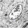
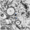
Representative EM photomicrographs show unstained preparations from aged human brains. (a) Ae micrograph (Case 1, 92-year-old, female) shows a neuron with a dense nucleolus within the nucleus. Numerous ribosomes and several mitochondria and ER structures can be visualized in the cytoplasm. Fibrils were visualized in the cytoplasm. (b) A micrograph (Case 2, 94-year-old, female) depicts an astrocyte filled with glial intermediate filaments on the right lower section of the image and a neuron's cytoplasm (left upper corner) containing a cluster of lipofuscins (LF) comprising electron dense rough-spherical intracytoplasmic structures, and a lipid body (LB). Neuropil can be appreciated between the astrocyte and the neuron. N: nucleus; M: mitochondrion; ER: endoplasmic reticulum; LB: lipid body; LF: lipofuscin; Np: neuropil, DB: dense body; Gfb: glial fibrils
Fig. 2
Fig. 2
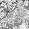
An unstained preparation showing high magnification of neuropil from an aged brain (94-year-old female, Case 2). M: mitochondrion; Ax: axon; AxT: axon terminal; D: dendrite; S: dendrite spine; Ast: astrocyte processes. Arrow: synapse
Fig. 3
Fig. 3
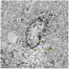
Low-magnification micrograph from a dementia case (90-year-old female, Case 6), labeled with PHF-1 antibody. pTau immunoreactivity can be seen throughout much of the neuronal cytoplasm with higher density in the peri-nuclear region and close to the rough ER and ribosome structures. N: nucleus, M: mitochondrion, ER: endoplasmic reticulum; G: Golgi body; LB: lipid body; Blue arrow: immunoreactivity of pTau; Yellow stars: consecutive serial micrographs were showed in Fig. 4a-d
Fig. 4
Fig. 4
a-d. Higher-magnification micrographs depict three consecutive serial sections at the location indicated in Fig. 3 with a yellow star. N: nucleus, M: mitochondrion, ER: endoplasmic reticulum; LB: lipid body. Blue arrows point to the appearance of some DAB immunoreactivity that was changed in the consecutive sections.
Fig. 5
Fig. 5
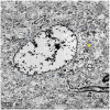
Low-magnification micrograph from a MCI case (86-year-old male, Case 5) labeled with MC-1 antibody. pTau immunoreactivity again appeared to mostly localize in ER structures and ribosomes. The neuron shown in the image had a invaginated nucleus and several small mitochondria. In addition, a cluster of lipid bodies (LB) and lipofuscins (LF) could be seen on the left side of the neuron. Blue arrows: immunoreactivity of pTau; N: nucleus, M: mitochondrion, ER: endoplasmic reticulum; LB: lipid body; LF: lipofuscin; Wide black arrow: nucleus invagination; Yellow stars: higher-magnification consecutive serial micrographs are depicted in Fig. 6
Fig. 6
Fig. 6
Six consecutive serial sections (from top left (#1) to bottom right (#6)) at the location indicated in Fig. 5 with a yellow star. N: nucleus; M: mitochondrion, ER: endoplasmic reticulum; Np: neuropil; Blue arrows pointed to the appearance of some DAB immunoreactivity changed in the consecutive sections
Fig. 7
Fig. 7
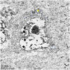
Low-magnification micrograph showing a cell from a MCI case (Case 5) stained with MC-1 antibody. The nucleus in the view was invaginated (black wide arrow) and ER structures were decorated with pTau immunoactivity. N: nucleus; M: mitochondrion; ER: endoplasmic reticulum; LB: lipid body; Blue arrows: immunoreactivity of pTau; Yellow star: consecutive serial micrographs were showed in Fig. 8
Fig. 8
Fig. 8
Six consecutive serial sections (from top left (#1) to bottom right (#6)) at the location indicated in Fig. 7 with a yellow star. N: nucleus; M: mitochondrion, ER: endoplasmic reticulum; LF: lipofuscin; LB: lipid body; Np: neuropil; Ax: axon; Blue arrows point to the location of some DAB immunoreactivity that was changed in the consecutive sections
Fig. 9
Fig. 9
Low-magnification micrograph showing a cell from Case 5 labelled with AT8 antibody. The nucleus showed invaginations (black wide arrows) and ER structures and ribosomes were decorated with pTau immunoactivity. N: nucleus; ER: endoplasmic reticulum; Blue arrows: immunoreactivity of pTau; Yellow star: location where consecutive serial micrographs are depicted in Fig. 10
Fig. 10
Fig. 10
Six consecutive serial sections (from top left (#1) to bottom right (#6)) at the location indicated in Fig. 9 with a yellow star. N: nucleus; M: mitochondrion, ER: endoplasmic reticulum; Blue arrows point to the appearance of some DAB immunoactivity that was changed in the consecutive sections
Fig. 11
Fig. 11
Electron micrograph from a 92-year-old female AD case (Case 1), which was immunogold labeled with MC-1 antibody. Gold particles were seen close to ribosomes and ER lumen. N: nucleus; ER: endoplasmic reticulum; rb: ribosome; M: mitochondrion
Fig. 12
Fig. 12
Immunogold labeled straight fibrils with PHF-1 antibody in a dementia case (84-year-old male, Case 7). Several large lipofuscins (LF) and lipid bodies (LB) were present in the neuron. N: nucleus; M: mitochondria; ER: endoplasmic reticulum; Fb: fibrils; LF: lipofuscin; LB: lipid body; Wide black arrow: nuclear invagination
Fig. 13
Fig. 13
High-magnification electron micrograph showing twisted Tau fibrils that were immunogold labeled with MC-1 antibody in a low AD pathology case (92-year-old female, Case 8). ER: endoplasmic reticulum; Fb: fibrils
Fig. 14
Fig. 14
Confocal images of pTau IF staining using MC-1 (a), AT8 (b), and PHF-1 (c) antibodies in the same case. In each collage, higher magnification images (scale bar = 10 μm) demonstrate the staining for various stages of NFTs. Low magnification image scale bar = 50 μm. These images were generated from an 81-year-old male dementia case (Case 9)
Fig. 15
Fig. 15
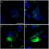
Representative confocal images of different Tau tangle development stage. The images depict an early stage granular pTau (a, b, pretangle), a fibrillary form of pTau (c, mature tangle), and a ghost tangle stage (d). These images were generated from an 86-year-old male MCI case (Case 5). This specific tissue section was IF stained using PHF-1 and Alexa 488 Goat anti-Mouse antibodies. Scale bar = 20 μm
Fig. 16
Fig. 16
Representative confocal images showing the interaction of granular pTau and ribosomes in the cytoplasm of pretangles (PHF-1 antibody). The upper panel images present the fluorescent labeling in a healthy neuron. The neuron was surrounded with several pTau positive neurites but no pTau was seen in the soma of the neuron. The lower panel images were taken from a pretangle neuron, in that, some granular pTau were overlaid with ribosomes (white arrow) and many of them seem to juxtapose with the ribosomal stains. Double immunofluorescence staining was carried out using ribosomal marker protein antibody RSP6 and the PHF-1 antibody on brain tissues obtained from a 81-year-old male dementia case (Case 9). Scale bar = 10 μm
Fig. 17
Fig. 17
3-D confocal image of the cytoplasm of a pretangle neuron double-labeled with pTau (AT8) and a ribosomal marker antibody. Double immunofluorescence staining was carried out with ribosomal marker protein antibody RSP6 and pTau antibody AT8 using brain tissue from a 81-year-old male dementia case (Case 9). The images depict the spatial relationship of granular pTau (green fluorescence) and the ribosomal marker (red fluorescence). Granular pTau was seen mostly juxtaposed with the ribosomal labeling but some granular pTau was overlaid with the ribosome marker (white arrow). Scale bar = 1 μm
Fig. 18
Fig. 18
Double immunofluorescent staining of pTau and ER marker Calnexin depicting a section from an 86-year-old female control case (Case 10). ER marker protein Calnexin and pTau (AT8 antibody) was labeled with Alexa Fluo 594 (red) and 488 (green), respectively. A pTau positive pretangle cell (white arrow) contained prominent granular pTau immunoreactivity, however, the pTau staining did not seem to completely overlap with the Calnexin stains. The yellow arrow points to a healthy neuronal cell that was not stained by AT8 antibody. Scale bar = 10 μm
Fig. 19
Fig. 19
Western blot analysis on the subcellular fractionation of human brain tissues. Brain tissues collected from the inferior parietal lobule were subjected to subcellular fractionation followed by Western blot analysis using either PHF-1 (pTau) or DA9 (non-phosphorylated Tau) antibody. Representative cases showed that pTau co-purified with and was enriched in ER and MAM fractions in the high Braak NFT stage cases (Case 12, 16). Lysate: post-nuclear lysate; pCyto: cytosol fraction; pMito: mitochondria fraction; MAM: mitochondria associated ER membrane fraction; ER: endoplasmic reticulum fraction; Brk: Braak NFT stage
Fig. 20
Fig. 20
Western blot analysis on the subcellular fractionation of wildtype (WT) and tauopathy mouse model (PS19Tg) brain tissues. Mice brain tissues were subjected to subcellular fractionation followed by Western blot analysis using either PHF-1 or DA9 antibody. While total Tau were present in both WT and PS19Tg mouse brain, pTau was seen only in ER and MAM fractions of the PS19Tg mouse brain. Lysate: post-nuclear lysate; pCyto: cytosol fraction; pMito: mitochondria fraction; MAM: mitochondria associated ER membrane fraction; ER: endoplasmic reticulum fraction

Similar articles

References

    1. Abisambra JF, Jinwal UK, Blair LJ, O’Leary JC 3rd, Li Q, Brady S, Wang L, Guidi CE, Zhang B, Nordhues BAet al et al (2013) Tau accumulation activates the unfolded protein response by impairing Endoplasmic reticulum-associated degradation. J Neurosci 33:9498–9507. 10.1523/JNEUROSCI.5397-12.2013 - PMC - PubMed
    1. Augustinack JC, Schneider A, Mandelkow EM, Hyman BT (2002) Specific Tau phosphorylation sites correlate with severity of neuronal cytopathology in alzheimer’s disease. Acta Neuropathol 103:26–35. 10.1007/s004010100423 - PubMed
    1. Bancher C, Brunner C, Lassmann H, Budka H, Jellinger K, Wiche G, Seitelberger F, Grundke-Iqbal I, Iqbal K, Wisniewski HM (1989) Accumulation of abnormally phosphorylated Tau precedes the formation of neurofibrillary tangles in alzheimer’s disease. Brain Res 477:90–99. 10.1016/0006-8993(89)91396-6 - PubMed
    1. Barnes DM (1985) Alz 50 recognizes an alzheimer’s protein. Sci (New York NY) 230:1260. 10.1126/science.4071048 - PubMed
    1. Besser L, Kukull W, Knopman DS, Chui H, Galasko D, Weintraub S, Jicha G, Carlsson C, Burns J, Quinn J et al (2018) Version 3 of the National Alzheimer’s Coordinating Center’s Uniform Data Set. Alzheimer Dis Assoc Disord 32: 351–358 10.1097/wad.0000000000000279 - PMC - PubMed

MeSH terms

LinkOut - more resources