. 2025 Jul 8;23(1):492.
doi: 10.1186/s12951-025-03574-w.

Quercetin nanoformulation-embedded hydrogel inhibits osteopontin mediated ferroptosis for intervertebral disc degeneration alleviation

Affiliations

Quercetin nanoformulation-embedded hydrogel inhibits osteopontin mediated ferroptosis for intervertebral disc degeneration alleviation

Jitian Li et al. J Nanobiotechnology. .

Abstract

Reactive oxygen species (ROS) play a pivotal role in multiple events during the progression of intervertebral disc degeneration (IDD). Hence, the precision treatment targets associated with ROS should be further explored to promote developing effective therapeutic strategies. In this study, by analyzing specimens from patients and RNA sequencing of ROS-induced human primary nucleus pulposus cells (NPCs), osteopontin (OPN) and ferroptosis were identified as critical molecular entities and cellular pathways implicated in ROS-mediated IDD. Subsequent animal models and cellular assays determined that ROS induced upregulation of OPN, which in turn triggered ferroptosis in NPCs and intervertebral discs, consequently leading to IDD. Building upon these findings, a comprehensive screening of molecular drug database revealed that quercetin, an antioxidant molecule compound, possesses the capacity to couple OPN, thereby mitigating OPN-induced ferroptosis and IDD. In addition, the compound of quercetin for targeting OPN was encapsulated in phenylboric acid modified dendrimer (G3-PBA) nanoparticles to improve its solubility, and then embedded in a ROS-degradable and injectable hydrogel, thereby achieving on-demand release of quercetin with the progression of IDD. Collectively, this study not only identified a novel therapeutic target, but also engineered an effective therapeutic strategy intended for the autonomous management of IDD.

Keywords: Ferroptosis; Injectable hydrogel; Intervertebral disc degeneration; Osteopontin; Quercetin.


Conflict of interest statement

Declarations. Competing interests: The authors declare no competing interests.

Figures
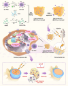
Fig. 1
Fig. 1
Schematic diagram of CMCS-PBA/PVA@G3-Que(quercetin@Gel) coupling with OPN to alleviate ferroptosis-related IDD
Fig. 2
Fig. 2
Oxidative stress promotes the degeneration of IVDs. (A) MRI analysis of patients with IDD according to Pfirrmann scores (the targeted IDD marked with red rectangle). (B) HE staining of human degenerative disc tissue (frozen section) with different degrees of IDD severity. (C, E) Quantitative DHE assay of ROS level in different IDD categories according to Pfirrmann scores; (D) HE and SOFG staining of rat IVD sections in the sham-surgery and H2O2-inoculation groups; (F) Histological grade analysis of rat IVD sections in the sham-surgery and H2O2-inoculation groups; (GH) Volcano plot and heat map in NPCs with or without ROS (H2O2) treatment; (I) Reactome enrichment analysis of differential gene expression in NPCs with or without ROS (H2O2) treatment; (J) Heat map of the top 50 differentially expressed genes in NPCs with or without ROS (H2O2) treatment; (KL) Protein–protein interaction (PPI) networks and hub gene analysis in NPCs with or without ROS (H2O2) treatment. (The data are shown as the mean ± SD. One-way ANOVA with Tukey’s multiple-comparison test was used for multiple group comparisons in E&F. n = 5 for each animal groups and n = 3 for RNA-seq analysis in each group. *P < 0.05, **P < 0.01, ***P < 0.001)
Fig. 3
Fig. 3
Ferroptosis is involved in oxidative stress–induced IDD. (A) MRI analysis of patients with IDD according to Pfirrmann scores (the targeted IDD marked with the red rectangle); (B) HE staining of human degenerative disc tissue (paraffin section) with different degrees of IDD severity; (C) Prussian Blue staining of iron deposition in degenerative disc tissue (Pfirrmann III–V); (D) Immunohistochemistry of ferroptosis-related proteins NRF2 and FTH1 in human degenerative disc tissue with different degrees of IDD severity; (E) ROS (H2O2) induced the degeneration and ferroptosis of NPCs in a dose-dependent manner as shown by western blot analysis; (F) Inhibition of ferroptosis in NPCs with Fer-1 alleviated the degeneration of NPCs. (G) TEM revealed the ferroptosis of NPCs with ROS (H2O2) and alleviation by Fer-1 (the mitochondria are indicated by white arrows); (H and I) Immunofluorescence staining of ferroptosis-related proteins GPX-4 and FTH1 in NPCs with ROS (H2O2) treatment and alleviation by Fer-1. (The data are shown as the mean ± SD. Two-way ANOVA with Dunnett’s multiple comparison test was used for E and Tukey’s test was used for F. n = 3 or 5 for each animal groups. All experiments were repeated for three times. *P < 0.05, **P < 0.01, ***P < 0.001, ****P < 0.0001, n.s.: no significance)
Fig. 4
Fig. 4
ROS-induced upregulation of OPN mediates ferroptosis and IDD. (A) MRI analysis of patients with IDD according to Pfirrmann scores (the targeted IDD is marked with the red rectangle); (B) HE staining and immunohistochemistry of OPN in human degenerative disc tissue with different degrees of IDD severity; (C) ROS induced the protein upregulation of OPN in NPCs in a dose- and time-dependent manner; (D) Immunofluorescence staining of ROS-induced overexpressed OPN in NPCs; (E) Immunohistochemistry and statistical analysis of OPN in rat IVDs after inoculation of H2O2; (F) OPN induced the degeneration and ferroptosis of NPCs in a dose-dependent manner as shown in western blot analysis; (G, I) HE and SOFG staining of rat IVDs after inoculation of low-dose (0.5 µg/per disc) and high-dose (1 µg/per disc) OPN, and histological grade analysis. (H, J) Immunohistochemistry and quantitative analysis of ferroptosis-related proteins FTH1 and NRF2 in rat IVDs from the sham-surgery and OPN-inoculation groups. (The data are shown as the mean ± SD. A two-sided Student’s t test for two-group analysis. One-way or Two-way ANOVA with Dunnett’s multiple comparison test was used for C&F, and with Tukey’s multiple-comparison test for I&J. The student’s t test was used for E. n = 3 or 5 for each animal groups. All experiments were repeated for three times. *P < 0.05, **P < 0.01, ***P < 0.001, ****P < 0.0001, n.s.: no significance)
Fig. 5
Fig. 5
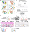
Quercetin inhibits ferroptosis of NPCs by coupling OPN. (A) Molecular docking structure diagram; (B) List of molecular docking binding energies; (C) Summary of SPR analysis between quercetin and OPN; (D) The binding of quercetin and OPN as determined by pulldown test; (E) The protein level of OPN after H2O2 stimulation with/without quercetin treatment in a dose-dependent manner, as shown by western blot analysis; (F) The protein level of ECM and ferroptosis-related protein after OPN (200 ng/mL, 24 h) stimulation with/without quercetin treatment in a dose-dependent manner, as shown by western blot analysis; (G, I) HE and SOFG staining of IVD sections of the rats in the sham-surgery, OPN (1 µg/per disc), and OPN with quercetin groups, and histological grade analysis; (H, J) Immunohistochemistry and quantitative analysis of FTH1 and NRF2 in the sham-surgery, OPN, and quercetin + OPN groups. (The data are shown as the mean ± SD. One-way or two-way ANOVA with Dunnett’s multiple comparison test for E&F and with Tukey’s multiple-comparison test was used for I&J. n = 3 or 5 for each animal groups. All experiments were repeated for three times. *P < 0.05, **P < 0.01, ***P < 0.001, ****P < 0.0001, n.s.: no significance)
Fig. 6
Fig. 6
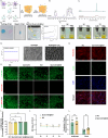
Characteristics and biocompatibility of G3-Que@Gel. (A) The process of G3-Que and CMCS-PBA/PVA hydrogel fabrication; (BC) 1H NMR spectrum of G3-PBA and CMCS-PBA polymer; (D) Hydrogel photography and integration of two broken hydrogel pieces; (E) Hydrogel injection using syringe needle and strain-dependent rheology of CMCS-PBA/PVA hydrogel with strain sweep from 0.1 to 2000% at angular frequency of 10 rad/second. (F) The morphology of hydrogel treated with H2O2 or PBS for different time. (G) Storage modulus evaluated by rheometer and surface morphology evaluated by SEM of the HA-PBA/PVA hydrogel treated with or without H2O2. (H and I) Live/dead staining of G3-Que@Gel cultured on NPCs at day 3. Live cells stained green, while dead cells stained red; (J) The survival rate of NPCs co-cultured with quercetin@Gel was detected by CCK-8 test on days 1, 3, and 5. (K and L) The toxicity of quercetin@Gel was examined by phalloidin and TUNEL staining of NPCs on day 3. (The data are shown as the mean ± SD. One-way or two-way ANOVA with Dunnett’s multiple comparison test was used for I-L. n = 3 or 5 for each animal groups. All experiments were repeated for three times.*P < 0.05, **P < 0.01, ***P < 0.001, ****P < 0.0001, n.s.: no significance)
Fig. 7
Fig. 7
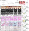
Quercetin hydrogel alleviates IDD. (A) Flowchart of experimental design and biomaterials administration; (B) Gross view of the quercetin hydrogel 1 h after the injection in a rat IVD; (CD) Gross examination and quantitative analysis of IVDs and nucleus pulposus area in the rats from the sham-surgery, IDD, IDD + gel, IDD + quercetin@Gel, and IDD + quercetin IP groups (Both the H2O2-induced and adjacent internal normal IVDs are marked with black ellipse); (EG) Radiological evaluation (MRI in T2-weighted imaging and micro-CT) and quantitative analysis of degenerative IVDs in each group. (HJ) HE and SOFG staining of degenerative IVDs in each group and histological quantitative analysis; (KM) Immunohistochemistry and quantitative analysis of collagen II and aggrecan in each group. (The data are shown as the mean ± SD. n = 3 ~ 5 for each group. One-way ANOVA with Dunnett’s multiple comparison test was used for multiple group comparisons at D-M. n = 3 or 5 for each animal groups.*P < 0.05, **P < 0.01, ***P < 0.001, ****P < 0.0001, n.s.: no significance)

References

    1. Battie MC, Videman T, Parent E. Lumbar disc degeneration: epidemiology and genetic influences. Spine (Phila Pa 1976). 2004;29(23):2679–90. - PubMed
    1. Teraguchi M, Yoshimura N, Hashizume H, Yamada H, Oka H, Minamide A, Nagata K, Ishimoto Y, Kagotani R, Kawaguchi H, et al. Progression, incidence, and risk factors for intervertebral disc degeneration in a longitudinal population-based cohort: the Wakayama spine study. Osteoarthritis Cartilage. 2017;25(7):1122–31. - PubMed
    1. Mohd Isa IL, Teoh SL, Mohd Nor NH, Mokhtar SA. Discogenic low back pain: anatomy, pathophysiology and treatments of intervertebral disc degeneration. Int J Mol Sci 2022;24(1). - PMC - PubMed
    1. Lin Y, Tang G, Jiao Y, Yuan Y, Zheng Y, Chen Y, Xiao J, Li C, Chen Z, Cao P. Propionibacterium acnes Induces Intervertebral Disc Degeneration by Promoting iNOS/NO and COX-2/PGE(2) Activation via the ROS-Dependent NF-kappaB Pathway. Oxid Med Cell Longev 2018;2018:3692752. - PMC - PubMed
    1. Chen Z, Jiao Y, Zhang Y, Wang Q, Wu W, Zheng J, Li J. G-Protein Coupled Receptor 35 Induces Intervertebral Disc Degeneration by Mediating the Influx of Calcium Ions and Upregulating Reactive Oxygen Species. Oxid Med Cell Longev 2022, 2022:5469220. - PMC - PubMed

LinkOut - more resources