. 2025 Jul 7;25(1):67.
doi: 10.1186/s12896-025-01001-4.

CDCP1 promotes the malignant phenotypes of nasopharyngeal carcinoma via the Wnt/β-catenin signaling pathway

Affiliations

CDCP1 promotes the malignant phenotypes of nasopharyngeal carcinoma via the Wnt/β-catenin signaling pathway

Guoliang Bie et al. BMC Biotechnol. .

Abstract

Background: CUB domain-containing protein 1 (CDCP1), a type I transmembrane glycoprotein, is abundantly expressed in various cancers. However, its role and mechanism in nasopharyngeal carcinoma (NPC) remain ambiguous.

Methods: The UALCAN and GEPIA databases were analyzed to explore CDCP1 expression and survival prognosis in head and neck squamous cell carcinoma (HNSC) patients. Fifteen pairs of NPC tissues and adjacent normal tissues were collected for CDCP1 expression analysis. CCK-8 assays, flow cytometry, and transwell assays were performed on NPC cell lines (C666-1, 5-8 F, and HONE-1). The impact of GSK-3β inhibitor LiCl on C666-1 cells after CDCP1 knockdown was investigated. A C666-1 xenograft model was established for in vivo validation.

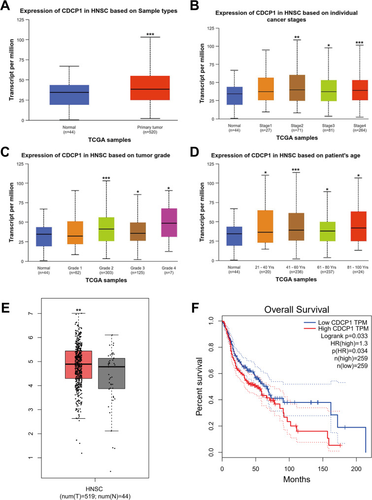
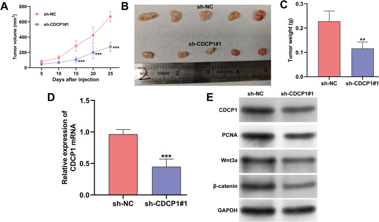
Results: CDCP1 was overexpressed in HNSC patients, and elevated CDCP1 correlated with poor survival. NPC tissues confirmed CDCP1 upregulation compared to normal tissues. CDCP1 knockdown in C666-1 and 5-8 F cells inhibited proliferation, migration, invasion, and promoted apoptosis, while LiCl partially reversed these effects. In vivo, CDCP1 silencing suppressed tumor growth, downregulated PCNA, Wnt3a, β-catenin, and p-GSK-3β, and upregulated cleaved caspase-3 and E-cadherin. CDCP1 overexpression in HONE-1 cells produced opposing effects.

Conclusions: In summary, CDCP1 promotes NPC progression via the Wnt/β-catenin pathway, suggesting its potential as a therapeutic target.

Clinical trial number: Not applicable.

Keywords: CDCP1; Nasopharyngeal carcinoma; Wnt/β-catenin signaling.


Conflict of interest statement

Declarations. Ethics approval and consent to participate: The samples utilized in this study were approved by the Medical Ethics Committee of the, Nanyang Central Hospital (Ethical Number: 202401007, Henan, China). Informed written consent was collected from all participants in accordance with the Declaration of Helsinki. The animal research was conducted in accordance with the National Institutes of Health Guidelines and was approved by the Animal Care and Use Committee of the Nanyang Institute of Technology (Henan, China). Consent for publication: Not applicable. Competing interests: The authors declare no competing interests.

Figures

Fig. 1
Fig. 1
Bioinformatic analysis of CDCP1 expression and survival prognosis in patients with HNSC. (A) The UALCAN database was employed to analyze the transcriptional expression of CDCP1 between HNSC tumors and normal tissues. (B) The difference in CDCP1 mRNA expression between HNSC tissues and normal tissues was evaluated based on individual cancer stages. (C) Analysis of CDCP1 mRNA expression difference according to tumor grade. (D) Examination of CDCP1 mRNA expression in relation to patient age. (E) The GEPIA database was utilized to analyze the transcriptional expression of CDCP1 between HNSC tumors and normal tissues. (F) Analysis of the TCGA database on the GEPIA database revealed the relationship between CDCP1 expression and the overall survival of HNSC patients. *p < 0.05, **p < 0.01, ***p < 0.001, compared with normal; HNSC, neck squamous cell carcinoma; GEPIA, Gene Expression Profiling Interactive Analysis
Fig. 2
Fig. 2
Validation of CDCP1 expression in clinical samples from NPC patients. (A) The mRNA expression of CDCP1 was assessed in 15 pairs of NPC tissues and adjacent normal tissues. (B) Representative four pairs of tumor and normal tissues were utilized to determine the protein level of CDCP1. ***p < 0.001 when compared with normal. The expression of CDCP1 at mRNA (C) and protein (D) levels was measured in three NPC cell lines (C666-1, 5–8 F, and HONE-1) and nasopharyngeal epithelial NP69 cell line. The data were shown as the mean ± SD. *p < 0.05, **p < 0.01, ***p < 0.001 when compared with NP69
Fig. 3
Fig. 3
CDCP1 regulated cell proliferation and apoptosis in NPC cells. (A-B) Quantitative real-time PCR and western blot analysis were employed to assess the expression of CDCP1 in C666-1 and 5–8 F cells at 48 h post-transfection with sh-CDCP1#1, sh-CDCP1#2, or sh-NC. (C) Confirmation of CDCP1 overexpression in HONE-1 cells in the presence of pcDNA3.1-CDCP1. (D) The CCK-8 assay was utilized to detect the cell proliferation rate in transfected C666-1, 5–8 F, and HONE-1 cells. Flow cytometry assay was used to determine cell apoptosis in transfected C666-1 (E), 5–8 F (F), and HONE-1 (G) cells. The data were shown as the mean ± SD. **p < 0.01, ***p < 0.001 when compared with sh-NC; ##p < 0.01, ###p < 0.001 when compared with pcDNA3.1
Fig. 4
Fig. 4
The influence of CDCP1 on the migration and invasion of NPC cells. C666-1 and 5–8 F cells were transfected with sh-CDCP1#1, and HONE-1 cells were transfected with pcDNA3.1-CDCP1 for 48 h. The transwell assay was employed to determine the migration and invasion capabilities of transfected C666-1 (A), 5–8 F (B), and HONE-1 (C) cells. The data were shown as the mean ± SD. **p < 0.01, ***p < 0.001 when compared with sh-NC; ##p < 0.01, ###p < 0.001 when compared with pcDNA3.1
Fig. 5
Fig. 5
Regulation of proliferation, apoptosis, and Wnt/β-catenin pathway-associated markers by CDCP1 in NPC cells. C666-1 and 5–8 F cells were transfected with sh-CDCP1#1, while HONE-1 cells were transfected with pcDNA3.1-CDCP1 for 48 h. The protein expression levels of PCNA, cleaved caspase-3, E-cadherin, Wnt3a, β-catenin, GSK-3β, and p-GSK-3β were determined in transfected C666-1 (A), 5–8 F (B), and HONE-1 (C) cells
Fig. 6
Fig. 6
Activation of Wnt/β-catenin pathway counteracted the consequences of CDCP1 knockdown in NPC cells. C666-1 cells were transfected with sh-CDCP1#1 and then treated with the GSK-3β inhibitor LiCl. (A) The cell proliferation rate was detected by the CCK-8 assay. (B) Flow cytometry assay was employed to assess cell apoptosis. (C) The transwell assay was utilized to determine the migration and invasion abilities of C666-1 cells. (D) Western blot analysis was performed to determine the protein expression levels of PCNA, E-cadherin, Wnt3a, and β-catenin. The data were shown as the mean ± SD. ***p < 0.001 when compared with sh-NC; ##p < 0.01, ###p < 0.001 when compared with sh-CDCP1#1
Fig. 7
Fig. 7
CDCP1 silencing repressed tumor growth of NPC in vivo. A xenograft subcutaneous model was established by stably infecting C666-1 cells with sh-CDCP1#1 or sh-NC, resulting in two groups with five mice each. (A) Tumor volumes in animal models were measured. (B) Representative images of tumors are presented. (C) Tumor weights were determined after euthanizing the mice. (D) Quantitative real-time PCR analysis validated the efficiency of CDCP1 knockdown by sh-CDCP1#1 in vivo. (E) Western blot analysis was employed to detect the protein expression of CDCP1, PCNA, Wnt3a, and β-catenin in tumors from the xenograft subcutaneous model. The data were shown as the mean ± SD. **p < 0.01, ***p < 0.001 when compared with sh-NC

Similar articles

References

    1. Qiao H, Tan XR, Li H, Li JY, Chen XZ, Li YQ, Li WF, Tang LL, Zhou GQ, Zhang Y, et al. Association of intratumoral microbiota with prognosis in patients with nasopharyngeal carcinoma from 2 hospitals in China. JAMA Oncol. 2022;8(9):1301–9. - PMC - PubMed
    1. Parlak S, Yazici G, Dolgun A, Ozgen B. The evolution of bone marrow signal changes at the skull base in nasopharyngeal carcinoma patients treated with radiation therapy. Radiol Med. 2021;126(6):818–26. - PubMed
    1. Feng G, Arima Y, Midorikawa K, Kobayashi H, Oikawa S, Zhao W, Zhang Z, Takeuchi K, Murata M. Knockdown of TFRC suppressed the progression of nasopharyngeal carcinoma by downregulating the PI3K/Akt/mTOR pathway. Cancer Cell Int. 2023;23(1):185. - PMC - PubMed
    1. Jiromaru R, Nakagawa T, Yasumatsu R. Advanced nasopharyngeal carcinoma: current and emerging treatment options. Cancer Manag Res. 2022;14:2681–9. - PMC - PubMed
    1. Zou W, Lei Y, Ding C, Xiao H, Wang S, Liang S, Luo W, Long Z, He S, Li Q, et al. The circadian gene ARNTL2 promotes nasopharyngeal carcinoma invasiveness and metastasis through suppressing AMOTL2-LATS-YAP pathway. Cell Death Dis. 2024;15(7):466. - PMC - PubMed

MeSH terms

LinkOut - more resources