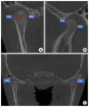
Assessment of upper airway and temporomandibular joint changes in growing patients with Class II Division 1 malocclusion, treated with the Twin Block appliance: a retrospective cone-beam computed tomography study
- PMID: 40624972
- PMCID: PMC12235306
- DOI: 10.7181/acfs.2024.0093
Assessment of upper airway and temporomandibular joint changes in growing patients with Class II Division 1 malocclusion, treated with the Twin Block appliance: a retrospective cone-beam computed tomography study
Abstract
Background: The Twin Block (TWB) appliance is widely employed for treating Class II malocclusion in children and adolescents. This study aimed to evaluate the three-dimensional treatment effects of the TWB on the upper airway and temporomandibular joint (TMJ), and to investigate the association between airway changes and TMJ alterations.
Methods: This retrospective study examined 24 cone-beam computed tomography (CBCT) scans from 12 patients (mean age, 12.30 ± 1.24 years) diagnosed with Class II Division 1 malocclusion and treated with the TWB appliance. CBCT scans were acquired both at pretreatment (T0) and posttreatment (T1). Romexis 6.2.1 imaging software was used to assess changes in the upper airway and TMJ. The paired t-test was used to compare the pretreatment and posttreatment measurements, while Pearson correlation coefficient analysis evaluated the relationship between the upper airway and TMJ measurements.
Results: A statistically significant increase was observed in the upper airway volume, condylar volume, and condylar dimensions after treatment. No significant correlation was detected between the upper airway and TMJ measurements at T0, T1, or in the net changes (T1-T0) during TWB therapy.
Conclusion: Growing patients treated with the TWB appliance demonstrated a statistically significant increase in upper airway volume. In addition, there was a significant increase in condylar volume, width, and length, with the condyle repositioned more anteriorly within the glenoid fossa. However, no statistically significant correlation was found between the TMJ and upper airway measurements.
Keywords: Adolescent; Cone-beam computed tomography; Functional orthodontic appliance; Malocclusion; Mandible; Temporomandibular joint.
Conflict of interest statement
No potential conflict of interest relevant to this article was reported.
Figures


Similar articles
-
Orthodontic treatment for prominent upper front teeth in children.Cochrane Database Syst Rev. 2007 Jul 18;(3):CD003452. doi: 10.1002/14651858.CD003452.pub2. Cochrane Database Syst Rev. 2007. PMID: 17636724
-
Positional and dimensional temporomandibular joint osseous changes in patients treated with the forsus fatigue resistant device: a non-randomized clinical trial.Clin Oral Investig. 2025 Aug 18;29(9):414. doi: 10.1007/s00784-025-06474-3. Clin Oral Investig. 2025. PMID: 40820194 Free PMC article. Clinical Trial.
-
Orthodontic treatment for prominent upper front teeth (Class II malocclusion) in children and adolescents.Cochrane Database Syst Rev. 2018 Mar 13;3(3):CD003452. doi: 10.1002/14651858.CD003452.pub4. Cochrane Database Syst Rev. 2018. PMID: 29534303 Free PMC article.
-
The relationship between the articular disc in magnetic resonance imaging and the condyle in cone beam computed tomography: A retrospective study.J Stomatol Oral Maxillofac Surg. 2024 Sep;125(12 Suppl 2):101940. doi: 10.1016/j.jormas.2024.101940. Epub 2024 Jun 8. J Stomatol Oral Maxillofac Surg. 2024. PMID: 38857693
-
Age and Gender-related Morphometric Assessment and Degenerative Changes of Temporomandibular Joint in Symptomatic Subjects and Controls using Cone Beam Computed Tomography (CBCT): A Comparative Analysis.Curr Med Imaging. 2024;20:e15734056248617. doi: 10.2174/0115734056248617231002110417. Curr Med Imaging. 2024. PMID: 38389339
References
-
- Ghafari JG, Macari AT. Component analysis of Class II, Division 1 discloses limitations for transfer to Class I phenotype. Semin Orthod. 2014;20:253–71.
-
- Bishara SE. Class II malocclusions: diagnostic and clinical considerations with and without treatment. Semin Orthod. 2006;12:11–24.
-
- Clark WJ. The Twin Block traction technique. Eur J Orthod. 1982;4:129–38. - PubMed
-
- Bjork A. Variations in the growth pattern of the human mandible: longitudinal radiographic study by the implant method. J Dent Res. 1963;42:400–11. - PubMed
-
- Koretsi V, Zymperdikas VF, Papageorgiou SN, Papadopoulos MA. Treatment effects of removable functional appliances in patients with Class II malocclusion: a systematic review and meta-analysis. Eur J Orthod. 2015;37:418–34. - PubMed
LinkOut - more resources
Full Text Sources

