Review
. 2025 Jun 23:13:1577192.
doi: 10.3389/fbioe.2025.1577192. eCollection 2025.

Recent advances in glycopeptide hydrogels: design, biological functions, and biomedical applications

Affiliations
Review

Recent advances in glycopeptide hydrogels: design, biological functions, and biomedical applications

Jinpeng Zhang et al. Front Bioeng Biotechnol. .

Abstract

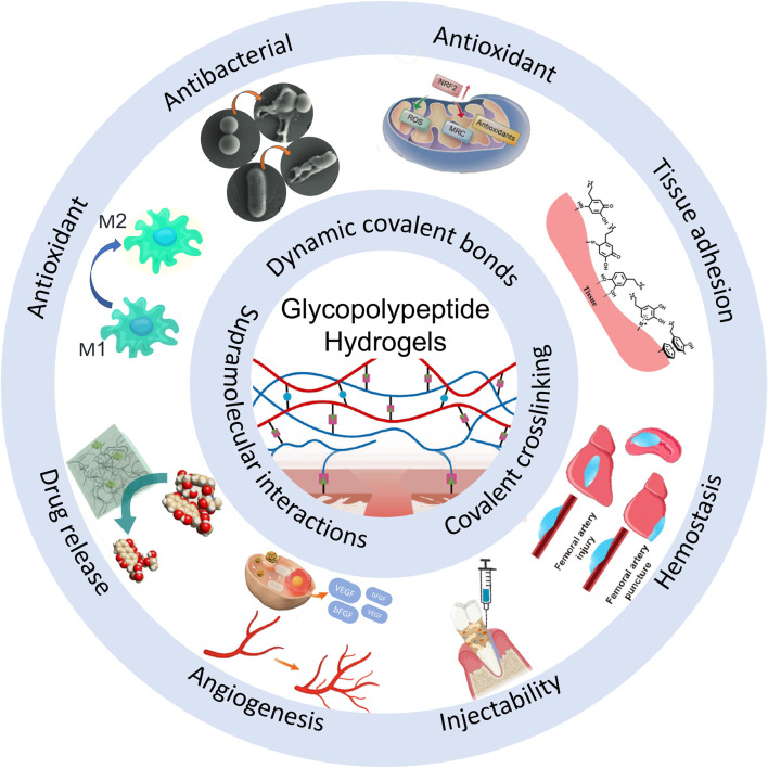
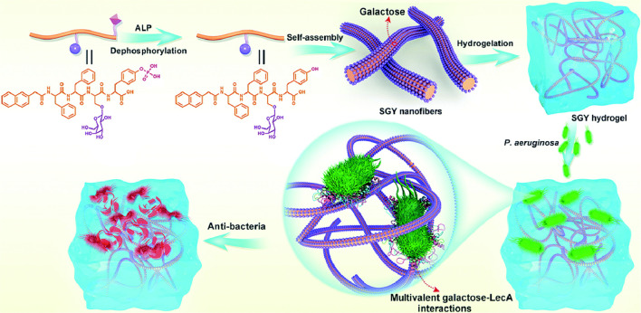
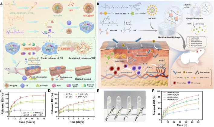
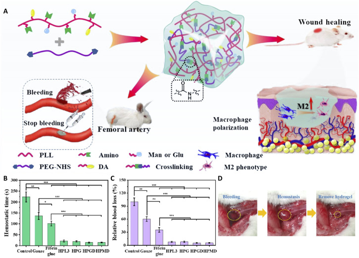
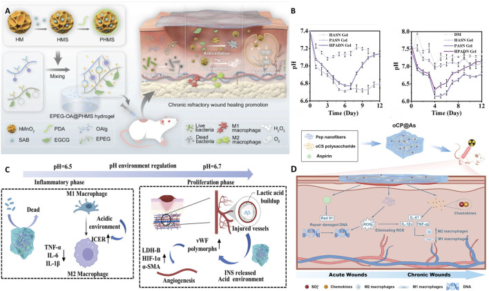
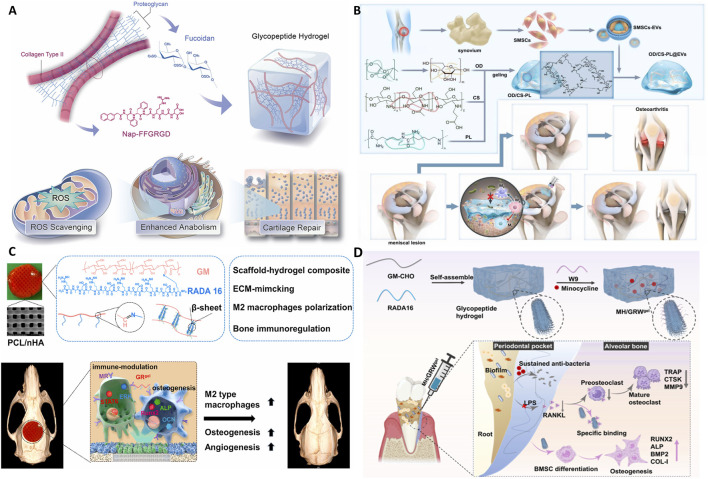
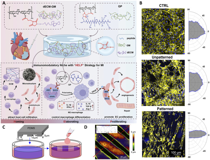
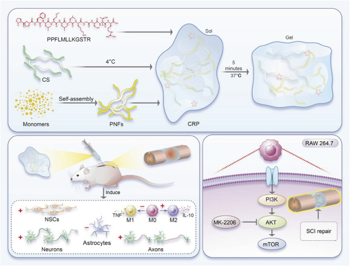
Glycopeptide hydrogels, biomaterials constructed from polysaccharides and peptides through dynamic covalent bonding and supramolecular interactions, mimic the structure and functions of the natural extracellular matrix. Their three-dimensional network structure endows them with remarkable mechanical resilience, self-healing capacity, and stimuli-responsive behavior, enabling diverse biomedical applications in tissue regeneration, wound healing, drug delivery, and antimicrobial therapies. This review comprehensively examines design principles for engineering glycopeptide hydrogels, encompassing biomolecular selection criteria and dynamic crosslinking methodologies. We analyze their multifunctional properties including antimicrobial efficacy, immunomodulation, antioxidant activity, tissue adhesion, and angiogenic potential, while highlighting smart drug release mechanisms. Applications in regenerative medicine are critically assessed, particularly in cutaneous wound healing, bone and cartilage reconstruction, myocardial repair, and neural regeneration. Finally, we delineate future directions to advance glycopeptide hydrogels, emphasizing functional sequence expansion of bioactive motifs, high-fidelity biomechanical mimicry of natural tissues, and precise simulation of organ-specific microenvironments for next-generation precision medicine.

Keywords: dynamic hydrogel; injectable hydrogel; self-assembling peptide hydrogel; supramolecular hydrogel; tissue engineering.


Conflict of interest statement

The authors declare that the research was conducted in the absence of any commercial or financial relationships that could be construed as a potential conflict of interest.

Figures

FIGURE 1
FIGURE 1
Schematic diagram of the biological functions of glycopeptide hydrogels. Antibacterial and angiogenesis (Wu et al., 2023), Copyright 2023. Reproduced with permission from John Wiley and Sons, Inc. Injectability (Zhao et al., 2024b), Copyright 2024. Reproduced with permission from the American Chemical Society. Hemostasis (Zhu et al., 2024), Copyright 2024. Reproduced with permission from the American Chemical Society. Drug release (Sakamoto et al., 2022), Copyright 2022. Reproduced with permission from the American Chemical Society. Antioxidant (Zhao et al., 2024a), Copyright 2023. Reproduced with permission from Elsevier B.V. Tissue adhesion (Teng et al., 2021), Copyright 2021. Reproduced with permission from John Wiley and Sons, Inc.
FIGURE 2
FIGURE 2
Schematic diagram of a multivalent sugar lectin-targeted antimicrobial hydrogel based on self-assembling peptides. (Liu et al., 2020), Copyright 2020. Reproduced with permission from the Royal Society of Chemistry.
FIGURE 3
FIGURE 3
Schematic illustrations and release profiles of smart glycopeptide hydrogels used for targeted drug delivery and wound healing. (A) Schematic illustration of the pH/ROS dual-responsive injectable glycopeptide hydrogel DS&MIC@MF. (B) Schematic illustration of the smart responsive hydrogel EPBA-PVA@MIC&AST. (C,D) Release kinetics of DS (C) and MF (D) from the DS&MIC@MF hydrogel under different conditions. (Wu et al., 2022), Copyright 2021. Reproduced with permission from Elsevier B.V. (E,F) pH/ROS dual-responsive behavior (E) and AST release kinetics (F) of the EPBA-PVA@MIC&AST hydrogel. (Deng et al., 2024), Copyright 2023. Reproduced with permission from Elsevier B.V. AST: astragaloside IV; DS: diclofenac sodium; EPBA: Phenylboronic acid grafted ε-Poly-L-lysine; MF: mangiferin; MIC: micelles; PVA: polyvinyl alcohol.
FIGURE 4
FIGURE 4
Application of a glycopeptide hydrogel in hemostasis. (A) Schematic illustration of the preparation and application of a biomimetic PEGylated glycopeptide hydrogel that modulated the inflammatory wound microenvironment to promote arterial hemostasis and wound healing. (B,C) In vivo hemostatic performance of the hydrogel in a rat femoral artery model. Relative blood loss (B) and hemostasis time (C) for different hydrogel formulations. (D) Representative photographs demonstrating the hemostatic effect of the hydrogel: (left) initial bleeding, (middle) hemostasis achieved with the hydrogel, and (right) hydrogel removal after hemostasis. (Teng et al., 2024), Copyright 2024. Reproduced with permission from the American Chemical Society.
FIGURE 5
FIGURE 5
Application of a glycopeptide hydrogel in the skin. (A) Schematic diagram of the preparation method and wound healing mechanism of the EPEG-OA@PHMS hydrogel. (Wu et al., 2023), Copyright 2023. Reproduced with permission from John Wiley and Sons, Inc. (B,C) Dynamic pH-regulated wound healing promoted by the HPADN hydrogel at different stages. HPADN hydrogel included HA modified with diacylhydrazine adipate (HA-ADH) or aldehyde (OHA), and dopa-modified poly(6-aminohexanoic acid) (PADA). (Liu et al., 2024), Copyright 2024. Reproduced with permission from Elsevier Science, Ltd. (D) Dual-phase regulatory strategy of the oCP@As hydrogel. (Guo et al., 2024), Copyright 2024. Reproduced with permission from the American Chemical Society. EPEG-OA: phenylboronic acid-grafted EPL, epigallocatechin-3-gallate and oxidized alginate; PHMS: resveratrol B-loaded, honeycomb-like MnO2 nanoparticles.
FIGURE 6
FIGURE 6
Application of glycopeptide hydrogels in bone defect repair. (A) Schematic diagram of the preparation and biological functions of the Nap-FFGRGD@Fucoidan hydrogel. (Zhao et al., 2024a), Copyright 2023. Reproduced with permission from Elsevier Science, Ltd. (B) Schematic diagram of the preparation and biological functions of the OD/CS-PL@EV hydrogel. (Huang et al., 2025), Copyright 2024. Reproduced with permission from Elsevier Science, Ltd. (C) Schematic diagram of the preparation and biological functions of PH@GR(gel). (Wang et al., 2022), Copyright 2022. Reproduced with permission from Elsevier Science, Ltd. (D) Schematic diagram of MH/GRW(gel). (Zhao et al., 2024b), Copyright 2024. Reproduced with permission from the American Chemical Society. CS: chitosan; EV: Extracellular vesicle; GRW: glucomannan, RADA16 and WP9QY peptide; PL: ε-Poly-L-lysine; OD: oxidized dextran; MH: minocycline hydrochloride; PH@GR(gel): glycopeptide hydrogel (GRgel) combined with a 3D-printed polycaprolactone and nano-hydroxyapatite (PCL/nHA) scaffold.
FIGURE 7
FIGURE 7
Application of glycopeptide hydrogels in myocardial tissue repair. (A) Schematic representation of the preparation and biological functions of dECM/GP. (Kong et al., 2023), Copyright 2023. Reproduced with permission from John Wiley and Sons, Inc. (B) Illustration of the elongation and alignment of iPSC-CMs cultured on control and Fmoc-FFGlcN6S hydrogel matrices. (C) Schematic of the micro-patterning process used. (D) Representative atomic force microscopy images of the micro-patterned hydrogels. (Castro et al., 2025), Copyright 2024. Reproduced with permission from Elsevier. dECM: decellularised extracellular matrix; GP: glycopeptide; iPSC-CMs: Induced pluripotent stem cell-derived cardiomyocytes.
FIGURE 8
FIGURE 8
Preparation of the CRP hydrogel and its application in spinal cord injury. (Sun et al., 2024), Copyright 2024. Reproduced with permission from John Wiley and Sons, Inc. CRP: chitosan, RADA16 and PPFLMLLKGSTR peptide.

Similar articles

References

    1. Aoki K., Saito H., Itzstein C., Ishiguro M., Shibata T., Blanque R., et al. (2006). A TNF receptor loop peptide mimic blocks RANK ligand–induced signaling, bone resorption, and bone loss. J. Clin. Invest 116 (6), 1525–1534. 10.1172/jci22513 - DOI - PMC - PubMed
    1. Apostolova E., Lukova P., Baldzhieva A., Katsarov P., Nikolova M., Iliev I., et al. (2020). Immunomodulatory and anti-inflammatory effects of fucoidan: a review. Polymers 12 (10), 2338. 10.3390/polym12102338 - DOI - PMC - PubMed
    1. Azuri I., Adler-Abramovich L., Gazit E., Hod O., Kronik L. (2014). Why are diphenylalanine-based peptide nanostructures so rigid? Insights from first principles calculations. J. Am. Chem. Soc. 136 (3), 963–969. 10.1021/ja408713x - DOI - PubMed
    1. Bai L., Wang T., Deng Q., Zheng W., Li X., Yang H., et al. (2024). Dual properties of pharmacological activities and preparation excipient: Bletilla striata polysaccharides. Int. J. Biol. Macromol. 254, 127643. 10.1016/j.ijbiomac.2023.127643 - DOI - PubMed
    1. Baranwal A., Kumar A., Priyadharshini A., Oggu G. S., Bhatnagar I., Srivastava A., et al. (2018). Chitosan: an undisputed bio-fabrication material for tissue engineering and bio-sensing applications. Int. J. Biol. Macromol. 110, 110–123. 10.1016/j.ijbiomac.2018.01.006 - DOI - PubMed

LinkOut - more resources