Review
. 2025 Jul 8;53(13):gkaf641.
doi: 10.1093/nar/gkaf641.

The ribosomal RNA transcription landscapes of Plasmodium falciparum and related apicomplexan parasites

Affiliations
Review

The ribosomal RNA transcription landscapes of Plasmodium falciparum and related apicomplexan parasites

Gunnar Mair et al. Nucleic Acids Res. .

Abstract

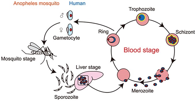
Ribosome biogenesis is essential for the rapid proliferation and life cycle transitions of Plasmodium falciparum, the causative agent of malaria. In eukaryotes, ribosomal RNA synthesis is carried out by RNA polymerase I (Pol I), highly specialized transcriptional machinery. This review provides a comparative analysis of Pol I transcription apparatus in yeast and humans, serving as a reference framework to examine its evolutionary divergence in P. falciparum and related apicomplexans and alveolates. Bioinformatic analyses revealed that some of these organisms lack any identifiable homologues or orthologs of several canonical eukaryotic transcription factors essential for Pol I-mediated transcription, including initiation factor RRN3, activator UBF, and all specific subunits of the promoter recognition complexes. Interestingly, the parasite retains core Pol I subunits, incorporating unique parasite-specific structural domains characterized through AI-based protein complex modeling of P. falciparum Pol I. These adaptations may compensate for the absence of traditional regulatory factors, enabling the parasite to employ distinct mechanisms for promoter recognition and transcription initiation. The substantial differences between parasite and host Pol I transcription machinery create potential targets for therapeutic intervention with parasite-specific elements representing potential drug targets. By integrating evolutionary, structural, and functional perspectives, this review advances our understanding of Pol I transcription in alveolates and its implications for the development of novel antimalarial strategies.


Conflict of interest statement

None declared.

Figures

Graphical Abstract
Graphical Abstract
Figure 1.
Figure 1.
The malaria parasite life cycle. A human malaria infection is initiated with the injection of sporozoites into the skin or circulatory system during the blood meal of a female Anopheles mosquito. The next port of call is the liver; here a single sporozoite produces thousands of daughter cells in an infected hepatocyte; these forms are called merozoites; infectious to the red blood cell, they are the cause of the clinical manifestations of malaria caused by the repeated infection of the erythrocytes. Transmission to the mosquito relies on the formation of male and female gametocytes within the red blood cell; following a blood meal by a female Anopheles gametocytes differentiate into mature male and female gametes and fuse to produce a zygote; the zygote develops into the motile ookinete, establishes a sessile oocyst, and in turn the sporozoites; they populate the salivary glands, which completes the life cycle [1, 2].
Figure 2.
Figure 2.
Ribosomal DNA loci in P. falciparum and P. berghei. The stage-specific expression patterns (A- and S-type) are indicated on the right with the corresponding gene identifiers. The five P. falciparum 5′ETS are from Fang et al. [37]; the two P. berghei 5′ETS (all shown as gray rectangles) are from van Spaendonk et al. [36]. The 2811 bp long noncoding RNA tru-lncRNA is indicated as a yellow rectangle. Seven noncoding RNAs are indicated as green rectangles. Arrows indicate direction of transcription. SRP, signal recognition particle; ncRNA, noncoding RNA; ITS, internal transcribed spacer; ETS, external transcribed spacer; and tRNA, transfer RNA. Drawn to scale from data available at plasmodb.org. The tru-lncRNA GenBank accession number: AY496275.
Figure 3.
Figure 3.
GC-content surrounding experimentally determined and proposed TSSs. TSSs are positioned at base position 101 in all sequences within the 200 base pair window. Plot generated at https://en.vectorbuildercom/tool/gc-content-calculator.html. The average GC-content is indicated by a line (https://www.ncbi.nlm.nih.gov/pmc/articles/PMC5857377/): P. berghei= 22.04%; P. falciparum= 19.34%.
Figure 4.
Figure 4.
The phylogenetic distribution of RNA Pol I transcription factors in selected eukaryotes. Core Pol I—core Pol I subunits; HD—subunits of Pol I-specific heterodimer; Stalk—subunits forming stalk domain; RRN3—Pol I associated transcription factor; SL1—subunits of human Selectivity factor 1; TBP—TATA-binding protein; CF—subunits of yeast Core Factor; UAF—subunits of yeast Upstream Activating Factor; and UBF—human Upstream Binding Factor. Phylogenetic tree of alveolates highlighting the relationships of selected apicomplexan parasites, chrompodellids, and the ciliate T. thermophila in comparison to Opisthokonta (H. sapiens and S. cerevisiae; adapted and generalized from Janouškovec at al. [62]). The headers refer to the components required for ribosomal DNA transcription identified in H. sapiens and yeast. Filled and empty squares indicate the presence and absence of proteins. The species refer to Babesia microti strain RI, Chromera velia CCMP2878, Cryptosporidium hominis isolate TU502_2012, Eimeria tenella Houghton 2021, Gregarina niphandrodes unknown strain, Haemoproteus tartakovskyi strain SISKIN1, Homo sapiens REF, P. falciparum 3D7 and P. berghei ANKA, Saccharomyces cerevisiae S288C, Sarcocystis neurona SN3, Tetrahymena thermophila (strain SB210), Theileria equi strain WA, Toxoplasma gondii ME49, and Vitrella brassicaformis CCMP3155, as indicated on https://orthomcl.org/orthomcl/app/ (release 6.21 of 7 May 2024).
Figure 5.
Figure 5.
Ribbon model of RNA Pol I highlighting specific subunits and domains. The “front view” looks along the incoming (“downstream”) DNA. Subunits not visible in the front view but present in the model are AC19, Rpb10, and Rpb12. Cyan spheres depict coordinated zinc atoms. Taken from Pilsl and Engel [63].
Figure 6.
Figure 6.
Comparison of the human (H.s.) and P. falciparum (P.f.) Pol I subunits with largest differences in size (RPA1, RPA2, RPA12, and RPA43). A, D, G, and H. Schematic outline of domain architecture of RPA1 (A), RPA2 (D), RPA12 (G), and RPA43 (H). The amino acid residue numbers at the domain boundaries are indicated. The largest amino acid insertions are marked by gray boxes. Ribbon diagram of RPA1 (B) and RPA2 (E). Domains are colored and color-coded as in panel (A) (RPA1) or as in panel (D) (RPA2). Inserts in P.f. subunits are in gray. A surface representation of RPA1 (C) and RPA2 (F). Domains are colored and color-coded as in panel (A) (RPA1) or as in panel (D) (RPA2). Inserts in P.f. subunits are in gray. Human subunits structure is from 7OB9; P.f. subunits are modeled in this work.
Figure 7.
Figure 7.
rRNA response in hmgb1 deletion mutants. FPMK values normalized for wild-type rRNA values. Data from Lu et al. [118].
Figure 8.
Figure 8.
Comparison of the predicted P. falciparum Pol I structure with its human counterpart. Left: Structure of elongating human Pol I (PDB 7OB9) shown in front, side (core module), back, and top views. Subunits are colored as follows: RPA1 (gray), RPA2 (wheat), RPAC1 (red), RPAC19 (yellow), RPABC1 (magenta), RPABC2 (hafnium), RPABC3 (green), RPABC4 (lemon), RPABC5 (density), RPA43 (slate), RPA12 (orange), RPA49 (light blue), and RPA34 (pink). Right: Structure predictions of P. falciparum Pol I presented in the same orientations. The composite model was constructed from predictions using different subunit combinations (see Methods). Conserved regions of P. falciparum Pol I are shown in gray, while large insertions relative to the human enzyme are highlighted in different colors: RPA1 (372–533: dark green, 616–675: green, 959–1085: dark violet, 1270–1770: cyan, 2302–2697: aquamarine blue), RPA2 (446–533: salmon), and RPA12 (1–225: orange). Bottom right of each view: Schematic overviews outline the orientations relative to the template. The Pol I core is shown in gray, the non-template DNA strand in medium blue, the template DNA strand in dark blue, and the synthesized RNA in red.

References

    1. Enomoto M, Kawazu S, Kawai S et al. Blockage of spontaneous Ca2+ oscillation causes cell death in intraerythrocitic Plasmodium falciparum. PLoS One. 2012; 7:e39499. 10.1371/journal.pone.0039499. - DOI - PMC - PubMed
    1. Sato S Plasmodium—a brief introduction to the parasites causing human malaria and their basic biology. J Physiol Anthropol. 2021; 40:1. - PMC - PubMed
    1. Baum J, Gilberger TW, Frischknecht F et al. Host-cell invasion by malaria parasites: insights from Plasmodium and Toxoplasma. Trends Parasitol. 2008; 24:557–63. 10.1016/j.pt.2008.08.006. - DOI - PubMed
    1. Lin J, Zhou D, Steitz TA et al. Ribosome-targeting antibiotics: modes of action, mechanisms of resistance, and implications for drug design. Annu Rev Biochem. 2018; 87:451–78. 10.1146/annurev-biochem-062917-011942. - DOI - PMC - PubMed
    1. Wilson DN Ribosome-targeting antibiotics and mechanisms of bacterial resistance. Nat Rev Micro. 2014; 12:35–48. 10.1038/nrmicro3155. - DOI - PubMed

Grants and funding

LinkOut - more resources