. 2025 Jul 29;10(7):e0006225.
doi: 10.1128/msphere.00062-25. Epub 2025 Jul 8.

LINC2781 enhances antiviral immunity against coxsackievirus B5 infection by activating the JAK-STAT pathway and blocking G3BP2-mediated STAT1 degradation

Affiliations

LINC2781 enhances antiviral immunity against coxsackievirus B5 infection by activating the JAK-STAT pathway and blocking G3BP2-mediated STAT1 degradation

Jiayu Zhang et al. mSphere. .

Abstract

Coxsackievirus B5 (CVB5) is a primary causative agent of hand, foot, and mouth disease (HFMD), and some cases are also associated with severe complications through invasion of the central nervous system, resulting in death. Currently, there are no specific antiviral drugs or effective vaccines available for CVB5. Long non-coding RNAs (lncRNAs) have been shown to play significant roles in various diseases. In our research, we identified a novel lncRNA, LINC2781, which is significantly upregulated during CVB5 infection of SH-SY5Y cells. Characteristic analysis showed that LINC2781 is mainly located in the cytoplasm of infected cells, with significantly higher expression in the intestines and the spleen of CVB5-infected mice. Functional studies revealed that LINC2781 activates the JAK-STAT pathway via STAT1, promoting the expression of interferon-stimulated genes (ISGs) and inhibiting CVB5 replication. Mechanistically, LINC2781 directly binds to GTPase-activating protein SH3 domain-binding protein 2 (G3BP2), preventing G3BP2-mediated degradation of STAT1 through ubiquitination. In vivo, LINC2781 was shown to reduce the susceptibility of BALB/c mice to viral infection and alleviate viral-induced damage in both the intestines and the spleen. Clinical samples further confirmed a strong correlation between the expression of LINC2781 and CVB5 infection. Our findings demonstrate that CVB5-induced LINC2781 enhances STAT1 activation by blocking the suppressive effects of G3BP2 on immune responses, providing a potential foundation for developing antiviral therapies targeting the lncRNA.

Importance: We investigate the role of lncRNA in virus-host interactions and identify a novel cytoplasmic lncRNA, LINC2781, whose expression is upregulated following CVB5 infection. LINC2781 specifically binds to G3BP2, preventing G3BP2 from degrading STAT1, thereby activating the JAK-STAT pathway, promoting the expression of ISGs, and ultimately inhibiting viral replication. Meanwhile, a strong correlation exists between the expression of LINC2781 and CVB5 infection in cells and clinical samples.

Keywords: LINC2781; GTPase-activating protein SH3 domain-binding protein 2 (G3BP2); STAT1; coxsackievirus B5 (CVB5); long non-coding RNAs (lncRNAs).


Conflict of interest statement

The authors declare no conflict of interest.

Figures

Fig 1
Fig 1
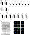
The characterization of LINC2781. (A) A schematic representation of the human LINC2781 transcribed from the XLOC_262866 gene; (B) SH-SY5Y and THP-1 cells were infected with CVB5 at increasing MOIs for 24 h or CVB5 (MOI = 1) for the indicated time intervals. The expression of LINC2781 was measured by RT-qPCR; (C) SH-SY5Y cells were transfected with increasing amounts of CVB5 RNA or plasmids (2 µg) encoding viral nonstructural proteins. The expression of LINC2781 was measured by RT-qPCR; (D) SH-SY5Y cells were infected with CVB5, CVA16, EV71, HSV-1, and RABV (MOI = 1) for 24 h. The expression of LINC2781 was measured by RT-qPCR; (E) SH-SY5Y cells were stimulated with different amounts of poly (I:C) or poly (dG:dC) for 24 h. The expression of LINC2781 was measured by RT-qPCR; (F) Various cell lines were infected with CVB5 (MOI = 1) for 24 h. The expression of LINC2781 was measured by RT-qPCR; (G) Three-day-old BALB/c mice were infected with CVB5 (20 LD50) for 10 days. The expression of LINC2781 was measured by RT-qPCR. (H) SH-SY5Y cells were treated with increasing amounts of IFN-β, IFN-γ, LPS, and TNF-α for 24 h. The expression of LINC2781 was measured by RT-qPCR. Biologically independent experiments (n = 3) were conducted, and all data were shown as mean ± SD. Student’s t-test was used to detect significant differences, with P ≤ 0.05 (*), P ≤ 0.01 (**), P ≤ 0.001 (***), and ns for no significant difference.
Fig 2
Fig 2
LINC2781 inhibits CVB5 replication. (A through C) LINC2781-overexpressing plasmid (LINC2781) or empty vector (pcDNA3.1) was transfected into SH-SY5Y cells, followed by infection with CVB5 (MOI = 1) at 24 h post-transfection. Cells and supernatants were harvested at 6, 12, or 24 h post-infection (hpi). The expression of CVB5 VP1 mRNA was measured by RT-qPCR (A), the expression of CVB5 VP1 protein was measured by western blotting (B), and the CVB5 titers were measured by TCID50 assay (C); (D through F) siLINC2781-1, siLINC2781-2, or empty vector (siNC) was transfected into SH-SY5Y cells, followed by infection with CVB5 (MOI = 1) at 24 h post-transfection. Cells and supernatants were harvested at 6, 12, or 24 hpi. The expression of CVB5 VP1 mRNA was measured by RT-qPCR (D), the expression of CVB5 VP1 protein was measured by western blotting (E), and the CVB5 titers were measured by TCID50 assay (F). Biologically independent experiments (n = 3) were conducted, and all data were shown as mean ± SD. Student’s t-test was used to detect significant differences, with P ≤ 0.05 (*), P ≤ 0.01 (**), P ≤ 0.001 (***), and ns for no significant difference. The band intensity of proteins was quantified, and the ratios of the target protein to α-Tubulin were shown.
Fig 3
Fig 3
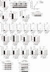
LINC2781 activates ISG expression and the JAK-STAT pathway via STAT1 activation. (A and B) LINC2781-overexpressing plasmid (LINC2781) or empty vector (pcDNA3.1) was transfected into SH-SY5Y cells, followed by infection with CVB5 (MOI = 1) at 24 h post-transfection. Cells and supernatants were harvested at 24 hpi. The secretion of IFN-I was measured by ELISA (A), and the expression of IFN-I and ISGs mRNA was measured by RT-qPCR (B); (C and D). siLINC2781-1, siLINC2781-2, or empty vector (siNC) was transfected into SH-SY5Y cells, followed by infection with CVB5 (MOI = 1) at 24 h post-transfection. Cells and supernatants were harvested at 24 hpi, the secretion of IFN-I was measured by ELISA, (C) and the expressions of IFN-I and ISGs mRNA were measured by RT-qPCR (D); (E) siLINC2781-1 or empty vector (siNC) was transfected into SH-SY5Y cells, followed by infection with CVB5 (MOI = 1) at 24 h post-transfection. Cells were harvested at 6, 12, and 24, and the expression of IFN/JAK-STAT pathway proteins was measured by western blotting; (F) LINC2781-overexpressing plasmid (LINC2781), siLINC2781-1, or empty vector was transfected into SH-SY5Y cells, followed by infection with CVB5 (MOI = 1) at 24 h post-transfection. Cells were harvested at 24 hpi, and the location and expression of IRF9 were measured by immunofluorescence. Biologically independent experiments (n = 3) were conducted, and all data were shown as mean ± SD. Student’s t-test was used to detect significant differences, with P ≤ 0.05 (*), P ≤ 0.01 (**), P ≤ 0.001 (***), and ns for no significant difference. The band intensity of proteins was quantified, and the ratios of the target protein to α-Tubulin were shown.
Fig 4
Fig 4
LINC2781 blocks G3BP2-mediated STAT1 degradation. (A) SH-SY5Y cells were infected with CVB5 (MOI = 1), then the cytoplasmic and nuclear fractions were collected and separated at 24 hpi. The expression of LINC2781 in different subcellular fractions was measured by RT-qPCR. GAPDH and U6 mRNA are, respectively, used as cytoplasmic and nuclear controls; (B) RNA pull-down analysis of LINC2781 binding to G3BP2. SH-SY5Y cells were infected with CVB5 (MOI = 1) and harvested at 24 hpi. The biotinylated LINC2781-positive or -negative strand was incubated with magnetic beads to obtain protein-RNA complexes. Complexes were then separated by 10% SDS-PAGE gel and analyzed by G3BP2 western blotting. (C) The binding between LINC2781 and G3BP2 was confirmed by RIP-qPCR. SH-SY5Y cells were infected with CVB5 (MOI = 1) and harvested at 24 hpi. Cell lysates were incubated with G3BP2 antibody (IgG as control) and magnetic beads to obtain the protein-RNA complexes. The total RNA was extracted from complexes, and the expression of LINC2781 was measured by RT-qPCR (%input). (D) pcDNA3.1-G3BP2-2Flag and pcDNA3.1-STAT1-6His were co-transfected into HEK293T cells for 24 h, and the Co-IP analysis was performed to determine the interaction between G3BP2 and STAT1. (E) pcDNA3.1-G3BP2-2Flag and pcDNA3.1-STAT1-6His were co-transfected into SH-SY5Y cells, followed by infection with CVB5 (MOI = 1) at 24 h post-transfection. The location and interaction between G3BP2 and STAT1 were conducted to immunofluorescence. (F) siG3BP2#3 or empty vector (siNC) was transfected into SH-SY5Y cells, followed by infection with CVB5 (MOI = 1) 24 h post-transfection. Cells were harvested at 24 hpi, and the expression of STAT1/p-STAT1 and G3BP2 was measured by western blotting. (G) pcDNA3.1-G3BP2 was transfected into SH-SY5Y cells, followed by infection with CVB5 (MOI = 1) 24 h pos-transfection. Cells were then treated for 48 h with autophagy inhibitor CQ, the caspase inhibitor Z-VAD-FMK, or the proteasome inhibitor MG132. The expression of STAT1/p-STAT1 and G3BP2 was measured by western blotting. (H) pcDNA3.1-G3BP2 and/or siPIAS1#2 were transfected into SH-SY5Y cells, followed by infection with CVB5 (MOI = 1) 24 h post-transfection. Immunoprecipitation analysis was performed to determine the ubiquitination of STAT1. (I) siLINC278-1 or empty vector (siNC) was transfected into SH-SY5Y cells, followed by infection with CVB5 (MOI = 1) 24 h post-transfection. Cells were harvested at 24 hpi, and Co-IP analysis was conducted to determine the interaction between G3BP2 and STAT1. Biologically independent experiments (n = 3) were conducted, and all data were shown as mean ± SD. Student’s t-test was used to detect significant differences, with P ≤ 0.01 (**) and ns for no significant difference. The band intensity of proteins was quantified, and the ratios of the target protein to α-Tubulin were shown.
Fig 5
Fig 5
LINC2781 relies on G3BP2 to exert antiviral effects. (A through E) siG3BP2#2, siG3BP2#3, or siNC was transfected into SH-SY5Y cells, followed by infection with CVB5 (MOI = 1) at 24 h post-transfection. Cells and supernatants were harvested at 24 hpi. The expression of CVB5 VP1 mRNA was measured by RT-qPCR (A). The expression of CVB5 VP1 protein was measured by western blotting (B). The CVB5 titers were measured by TCID50 assay (C). The secretion of IFN-I was measured by ELISA (D). The expression of IFN-I and vital ISGs mRNA was measured by RT-qPCR (E). F-I. siG3BP2#2, siG3BP2#3, and siLINC2781-1 were co-transfected into SH-SY5Y cells, followed by infection with CVB5 (MOI = 1) at 24 h post-transfection. Cells and supernatants were harvested at 24 hpi. The expression of CVB5 VP1 was measured by RT-qPCR and western blotting (F). The expression of IFN-I was measured by ELISA and RT-qPCR (G). The expression of vital ISGs mRNA was measured by RT-qPCR (H). The expressions of STAT1/p-STAT1 and IRF9 were measured by western blotting (I). (J) LINC2781-overexpressing plasmid (LINC2781) or empty vector (pcDNA3.1) was transfected into G3BP2 KO cells, followed by infection with CVB5 (MOI = 1) at 24 h post-transfection. Cells and supernatants were harvested at 24 hpi. The expression of STAT1/p-STAT1, CVB5 VP1, and G3BP2 was measured by western blotting. Biologically independent experiments (n = 3) were conducted, and all data were shown as mean ± SD. Student’s t-test was used to detect significant differences, with P ≤ 0.05 (*), P ≤ 0.01 (**), P ≤ 0.001 (***), and ns for no significant difference. The band intensity of proteins was quantified, and the ratios of the target protein to α-Tubulin were shown.
Fig 6
Fig 6
The 5’-terminal of LINC2781 is the main functional site. (A) The secondary structure of LINC2781 was analyzed by RNA-fold. Base pairing probabilities are color-coded on a scale from 0 (blue) to 1 (red). (B-D) LINC2781-overexpressing plasmid (LINC2781), its truncated functional domain plasmids (LINC2781-1, LINC2781-2, and LINC2781-3) or an empty vector (pcDNA3.1) was transfected into SH-SY5Y cells, followed by infection with CVB5 (MOI = 1) 24 h post-transfection. Cell lysates were incubated with the G3BP2 antibody (IgG as control) and magnetic beads to obtain the protein-RNA complexes. The total RNA was extracted from complexes, and the expression of LINC2781 was measured by RT-qPCR (%input). (B) The expression of STAT1/p-STAT1, G3BP2, and CVB5 VP1 was measured by western blotting (C). The expression of CVB5 VP1, IFN-β, OASL, and ISG15 mRNAs was measured by RT-qPCR (D). Biologically independent experiments (n = 3) were conducted, and all data were shown as mean ± SD. Student’s t-test was used to detect significant differences, with P ≤ 0.05 (*), P ≤ 0.01 (**), P ≤ 0.001 (***), and ns for no significant difference. The band intensity of proteins was quantified, and the ratios of the target protein to α-Tubulin were shown.
Fig 7
Fig 7
The role of LINC2781 in vivo. (A). Schematic diagram of the mice models. Three-day-old BALB/c mice were injected intraperitoneally with AAV2/9-LINC2781 (5 × 10¹¹ genomic copies) or AAV2/9 (2 × 10¹¹ genomic copies) thrice. Fourteen days later, the mice were infected with CVB5 (20 LD50) by intraperitoneal injection. Samples were collected 10 days post-infection. (B) Appearance characteristics of the mice. (C) Changes in mice body weight. (D) The expression of CVB5 was measured by TCID50 assay and RT-qPCR. (E) Pathological symptoms in the spleen and intestines were assessed by HE staining, and the expression of G3BP2 in the spleen and intestines was measured by IHC. (F) The expression of CVB5 VP1, IFN-β, OASL, and ISG15 mRNAs in the spleen and intestines was measured by RT-qPCR. (G) The expression of LINC2781 in clinical samples was measured by RT-qPCR. Six BALB/c mice were used for the experimental group, and all data were shown as mean ± SD. Student’s t-test was used to detect significant differences, with P ≤ 0.05 (*), P ≤ 0.01 (**), and ns for no significant difference.

Similar articles

References

    1. Machado RS, Tavares FN, Sousa IP. 2024. Global landscape of coxsackieviruses in human health. Virus Res 344:199367. doi: 10.1016/j.virusres.2024.199367 - DOI - PMC - PubMed
    1. Zhang X, Zhang Y, Li H, Liu L. 2022. Hand-foot-and-mouth disease-associated enterovirus and the development of multivalent HFMD vaccines. IJMS 24:169. doi: 10.3390/ijms24010169 - DOI - PMC - PubMed
    1. Di Prinzio A, Bastard DP, Torre AC, Mazzuoccolo LD. 2022. Hand, foot, and mouth disease in adults caused by coxsackievirus B1-B6. An Bras Dermatol 97:321–325. doi: 10.1016/j.abd.2021.03.012 - DOI - PMC - PubMed
    1. Andino R, Kirkegaard K, Macadam A, Racaniello VR, Rosenfeld AB. 2023. The picornaviridae family: knowledge gaps, animal models, countermeasures, and prototype pathogens. J Infect Dis 228:S427–S445. doi: 10.1093/infdis/jiac426 - DOI - PubMed
    1. Mattick JS, Amaral PP, Carninci P, Carpenter S, Chang HY, Chen L-L, Chen R, Dean C, Dinger ME, Fitzgerald KA, et al. 2023. Long non-coding RNAs: definitions, functions, challenges and recommendations. Nat Rev Mol Cell Biol 24:430–447. doi: 10.1038/s41580-022-00566-8 - DOI - PMC - PubMed

LinkOut - more resources