. 2025 Nov 3;31(21):4543-4556.
doi: 10.1158/1078-0432.CCR-25-0950.

Human Kallikrein 2: A Novel Lineage-Specific Surface Target in Prostate Cancer

Affiliations

Human Kallikrein 2: A Novel Lineage-Specific Surface Target in Prostate Cancer

Fei Shen et al. Clin Cancer Res. .

Abstract

Purpose: Targeted therapies for metastatic prostate cancer are limited, highlighting the need for novel drug targets and mechanisms of action (MoA). Human kallikrein 2 (KLK2) is a prostate-specific antigen expressed across the prostate cancer disease continuum. However, it was not recognized as a therapeutic target for prostate cancer in the past due to limited evidence of its cell surface expression. In this study, we systematically characterized KLK2 expression in prostate cancer, confirmed its cell surface expression, and demonstrated the preclinical efficacy of three KLK2-targeting therapeutics with distinct MoA.

Experimental design: The KLK2 expression profile in different stages of prostate cancer and its cell surface expression were confirmed by IHC and multiplex immunofluorescent staining. The preclinical efficacy of three KLK2-targeting therapeutics was characterized using in vitro prostate cancer cell lines, patient-derived material, and in vivo xenograft mouse models.

Results: KLK2 was found to be robustly and homogeneously expressed in localized prostate cancer and metastatic hormone-sensitive prostate cancer, whereas some heterogeneity was observed in the visceral lesions of metastatic castration-resistant prostate cancer. KLK2 expression was more specific than that of other prostate cancer target antigens. Although KLK2 is traditionally described as a secreted protease, our results demonstrated its cell surface expression in both prostate cancer cell lines and patient-derived tumors. Notably, targeting KLK2 with three different MoAs, including bispecific T-cell redirector, targeted α-radioligand, and autologous chimeric antigen receptor T cells, showed potent in vitro activity and robust in vivo tumor control.

Conclusions: Our study establishes KLK2 as a highly prostate-specific cell surface target. Targeting KLK2 with various MoAs represents novel therapeutic approaches for advanced prostate cancer. See related commentary by Blinka and Yu, p. 4393.


Conflict of interest statement

F. Shen reports other support from Johnson & Johnson outside the submitted work. R. Smith reports other support from Johnson & Johnson outside the submitted work. T. McDevitt reports other support from Johnson & Johnson outside the submitted work. K. Menard reports other support from Johnson & Johnson outside the submitted work. S. Tian reports other support from Johnson & Johnson outside the submitted work. G. Chu reports other support from Johnson & Johnson outside the submitted work. R. Chaudhary reports other support from Johnson & Johnson outside the submitted work. J. McCann reports other support from Johnson & Johnson outside the submitted work. H. Oyer reports other support from Johnson & Johnson Innovative Medicine outside the submitted work. S.C. Wang reports other support from Johnson & Johnson outside the submitted work. P. Francis reports other support from Johnson & Johnson outside the submitted work. W.K. Kelly reports grants from Janssen during the conduct of the study as well as nonfinancial support from Janssen and Amgen outside the submitted work. C.G. Drake reports other support from Johnson & Johnson Innovative Medicine outside the submitted work. No disclosures were reported by the other author.

Figures
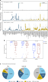
Figure 1.
Figure 1.
KLK2 expression profile. A, Box plot analysis of the KLK2, PSMA, and STEAP1 relative expression in normal human tissue types (n = 43) and cancer types (n = 68). The box refers to the quartile distribution (25%–75%) range, with the median shown as a black horizontal line. In addition, the 95% range and individual outlier samples are shown. B, KLK2 and PSMA protein expression in different stages of prostate cancer [LPC (n = 100), mHSPC (n = 100), and mCRPC (n = 45)] by IHC with semiquantitative pathologist reporting of the percentage of tumor showing any staining. The box refers to the IQR (25%–75% of samples), with the median shown as a horizontal line in the middle. In addition, whisker length extends to 1.5 × IQR on both sides of the IQR, and individual outlier samples are shown. C, Proportions of bone, lymph node, and soft tissue lesions showing coexpression of KLK2 and PSMA, as evaluated by mIF. The lesion is considered positive when ≥50% of cells are stained positive by mIF. AML, acute myeloid leukemia; B-ALL, B-cell acute lymphoblastic leukemia; B-CLL, B-cell chronic lymphocytic leukemia; CML, chronic myeloid leukemia; GBM, glioblastoma multiforme; GIST, gastrointestinal stromal tumor; MALT, mucosa-associated lymphoid tissue; NOS, not otherwise specified; PNS, peripheral nervous system; T-ALL, T-cell acute lymphoblastic leukemia.
Figure 2.
Figure 2.
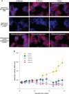
Cell surface expression of KLK2. A, FACS staining of KLK2 in VCaP cells. A representative FACS plot from n = 3 repeated experiments is shown. B, Workflow and FACS staining of KLK2 in freshly dissociated mCRPC tumor cells. Representative FACS plot is shown; n = 3. C, Confocal imaging of KLK2 surface expression in VCaP cells. A representative IF images are shown from n = 3 repeated experiments: KLK2 stain (green), EpCAM cell surface stain (red), and Hoechst DNA stain (blue).
Figure 3.
Figure 3.
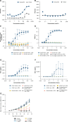
In vitro and in vivo characterization of KLK2 × CD3 in preclinical models. A and B, Binding of KLK2 × CD3 to (A) VCaP (KLK2+) and (B) DU145 (KLK2) cells. A representative dose–response binding curve is shown; n = 5. C–F, Evaluation of KLK2 × CD3 on (C) T cell–mediated cytotoxicity, (D) T-cell activation, and (E and F) TNF-α and IFN-γ cytokine release in vitro, in PBMC from five healthy donors. Mean values ± SEM are shown. n = 5. G, T cell–engrafted NSG mice bearing established VCaP tumors were intraperitoneally dosed with KLK2 × CD3 at 0.2 mg/kg, 1 mg/kg, 2.5 mg/kg, 5 mg/kg, and 15 mg/kg, compared with control (eight total doses). Tumor volume was measured twice weekly, and the results are presented as the mean tumor volume ± SEM for each group (n = 8 per group).
Figure 4.
Figure 4.
Preclinical characterization of KLK2-targeted RLT. A, Cell binding and internalization of the anti-KLK2 antibody in VCaP cells assessed by confocal imaging. Cell surface binding of the anti-KLK2 antibody was detected at 60 minutes with intracellular puncta formation observed at 3 hours, indicating antibody internalization. A representative confocal image is shown; n = 3. B, Treatment with a single i.v. dose of 225Ac-KLK2 in an established prostate xenograft model at 50, 100, 250, and 500 nCi, compared with control. Tumor volume was measured twice weekly, and the results are presented as the mean tumor volume ± SEM for each group (n = 10 per group).
Figure 5.
Figure 5.
Preclinical characterization of KLK2-targeted CAR T cells. A, KLK2-targeted CAR T cells exhibit cytotoxicity when cocultured with KLK2+ but not with KLK2 prostate cancer cell lines. The mean percentage of tumor lysis ± SEM is shown. A representative dose–response curve is shown; n = 3. B, In vivo i.v. treatment with KLK2-targeted CAR T cells (1 × 10⁶, 5 × 10⁶, and 10 × 106 CAR T cells) on established VCaP human prostate xenografts. Tumor volume was measured twice weekly, and the results are presented as the mean tumor volume ± SEM for each group (n = 10 per group).

References

    1. Siegel RL, Giaquinto AN, Jemal A. Cancer statistics, 2024. CA Cancer J Clin 2024;74:12–49. - PubMed
    1. National Cancer Institute . Cancer stat facts: prostate cancer. National Cancer Institute Surveillance, Epidemiology, and End Results (SEER) Program (2024). [cited 2024 Jul 10]. Available from:https://seer.cancer.gov/statfacts/html/prost.html.
    1. Sekhoacha M, Riet K, Motloung P, Gumenku L, Adegoke A, Mashele S. Prostate cancer review: genetics, diagnosis, treatment options, and alternative approaches. Molecules 2022;27:5730. - PMC - PubMed
    1. Mohile SG, Mustian K, Bylow K, Hall W, Dale W. Management of complications of androgen deprivation therapy in the older man. Crit Rev Oncol Hematol 2009;70:235–55. - PMC - PubMed
    1. Le TK, Duong QH, Baylot V, Fargette C, Baboudjian M, Colleaux L, et al. Castration-resistant prostate cancer: from uncovered resistance mechanisms to current treatments. Cancers (Basel) 2023;15:5047. - PMC - PubMed