. 2025 Jul 8;36(1):58.
doi: 10.1007/s10856-025-06908-0.

Ameliorative role of silver nanoparticles incorporated with chitosan solution and leukocyte platelet-rich fibrin scaffold during colon anastomosis in rabbits

Affiliations

Ameliorative role of silver nanoparticles incorporated with chitosan solution and leukocyte platelet-rich fibrin scaffold during colon anastomosis in rabbits

Mohammed Albahrawy et al. J Mater Sci Mater Med. .

Abstract

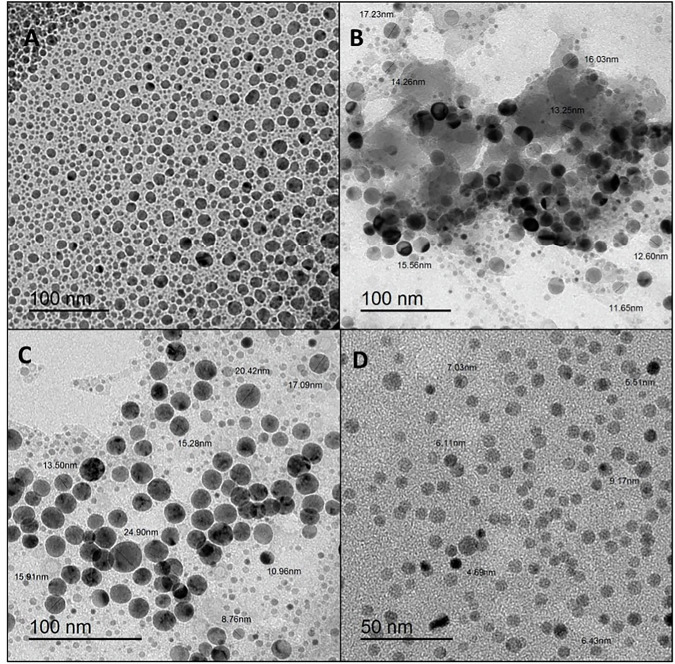
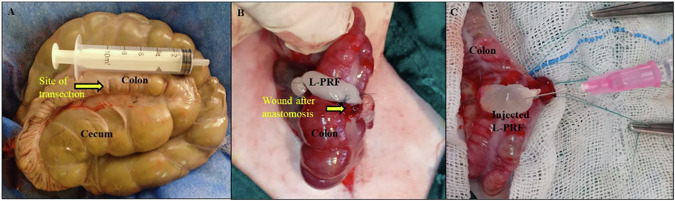
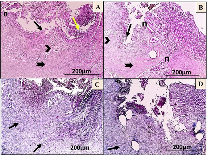
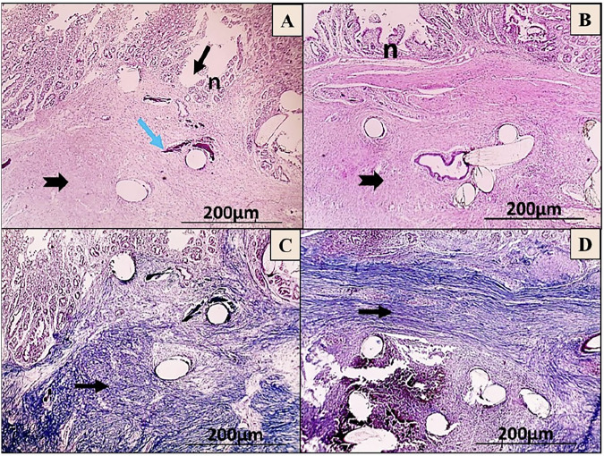
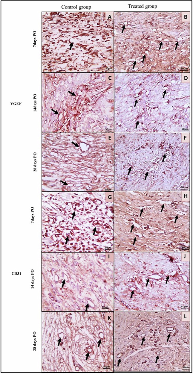
This research aimed to detect the experimental value of silver nanoparticles (AgNPs) loaded with chitosan (CS) and leukocyte platelet-rich fibrin (L-PRF) in promoting the healing process of the anastomotic colon in rabbits. Forty-two healthy male white New Zealand rabbits underwent total transection and anastomosis of the ascending colon. The rabbits were then randomly divided and equally allocated into two groups (n = 21): the control (C) group, in which the transected colon was traditionally anastomosed with simple interrupted sutures, and the treated (T) group, in which the colon was traditionally anastomosed and completely wrapped by a L-PRF scaffold that was inoculated with 1 ml of AgNPs-CS solution. Clinical macroscopic observations of rabbits and pain scale scores were evaluated postoperatively on the 7th,14th, and 28th days. Additionally, seven rabbits from each group were euthanized at each time point to assess bursting pressure, radiographic stenotic degree, histopathological findings, and immunohistochemical examination. The anastomotic colon in the T-group revealed a significant improvement in the overall health status of rabbits, as well as mechanical bursting pressure scores. Pain scores and radiographic stenotic degree were significantly lower in the T-group. Moreover, the histopathological examination of the anastomotic colon in the T-group showed an increased collagen deposition in the submucosa, proper re-epithelialization of the mucosa, higher angiogenesis, reduced adhesions, and significantly higher histopathological healing scores than the C-group. The AgNPs-CS/L-PRF scaffold successfully reduced complications following colon anastomosis and potentially ameliorated the healing process of the anastomotic colon in the experimental rabbits.


Conflict of interest statement

Compliance with ethical standards. Conflict of interest: The authors declare no competing interests. Ethics approval: All experiments were performed following relevant guidelines and regulations. This study was approved by the Ethics Committee of Mansoura University Animal Care and Use Committee with the code number MU-ACUC (VM.PhD.22.11.6). All procedures in this study were performed following ARRIVE guidelines and carried out in accordance with the U.K. Animals (Scientific Procedures) Act 1986 and associated guidelines.

Figures

None
Schematic cartoon of the experimental study. The authors of the manuscript designed it. (Created with Biorender.com with permission).
Fig. 1
Fig. 1
Transmission electron microscopy (TEM) images of different sizes of AgNPs-CS at various magnifications (AC) 100 nm, and D 50 nm
Fig. 2
Fig. 2
A A representative SAED pattern of AgNPs-CS. B UV-visible spectroscopic measurement of AgNPs-CS. C Zeta potential measurements for AgNPs in polymer matrix CS
Fig. 3
Fig. 3
A Site of ascending colon transection. B L-PRF clot fixation on anastomotic colon site (yellow arrow). C L-PRF after CS-AgNPs injection
Fig. 4
Fig. 4
Bristol Rabbit Pain Scale (BRPS) between the control (C) groups and the AgNPs-CS/L-PRF treated (T) group the day before surgery (day 0) and on the 7th, 14th, and 28th days post-operations
Fig. 5
Fig. 5
The Rabbit Facial expression according to the Bristol Rabbit Pain Scale (BRPS). A–C Rabbits in the C-group were dull, hunched backs, their Squinting, semi-closed eyes, nasal flare, and their ears directed backward. D–F Rabbits in the T-group were awake, lying with semi-closed eyes, and their ears slightly moved towards sounds
Fig. 6
Fig. 6
Adhesion grades in the control (C) group and the AgNPs-CS/L-PRF treated (T) group (D-F) groups. A Grade 2, soft, vascularized adhesion (stars) between colon anastomosis area (yellow arrow) and abdominal wall incision and cecum. B Grade 3, vascularized, solid (star) adhesion. C Grade 4, vascularized, solid, conglomerate adhesion (star) between the anastomotic line (yellow arrow), small intestine, and cecum. D, E Grade zero adhesion, F grade 1 easy, detached filamentous adhesion (star) between the anastomotic line (yellow arrow) and the cecum
Fig. 7
Fig. 7
The degrees of stenosis of the anastomotic line (black dots) were evaluated post-contrast radiographic scanning in the control (C) group and the AgNPs-CS/L-L-PRF treated (T) group at the different time points postoperative
Fig. 8
Fig. 8
Microscopic images of anastomosis wounds in the control (C) group (A, C) and AgNPs-CS/L-PRF treated (T) group (B, D) at day 7 post-operation. The wound shows extensive polymorph nuclear cell infiltration (yellow arrows) throughout the layers of the colon, deep ulceration that reaches adventitia (black arrow), and minimal granulation tissue formation (black arrowhead) (A). Black arrowhead-shaped granulation tissue development with noticeable re-epithelialization (n) (B) (HE; X:40). As well, the wound displays scattered bluish stained fibrils of collagen (black arrow) (C), fine bluish stained collagen forming thin bundles (black arrow) (D) (MT; X:40)
Fig. 9
Fig. 9
Microscopic images of anastomosis wounds in the control (C) group (A, C) and AgNPs-CS/L-PRF treatment group (B, D) at day 14 post-operation. The wound shows enhanced deposition of collagen (thick black arrow) in the submucosa, along with the formation of granulation tissue (black arrowhead), substantial infiltration of PMN (yellow arrow), and ulceration that reaches the Submucosa (black arrow) (A). Focal ulceration with increased re-epithelialization (n) that reaches the lamina propria (black arrow, thin one), greater collagen deposition (thick black arrow) is observed with granulation tissue formation (black arrowhead) (B) (HE; X:40). In addition, less bluish-stained collagen fibrils (black arrows) were found in the lamina propria and submucosa of the wounds (C), Higher and denser bluish bundles of collagen deposition (black arrow) appear in submucosa (D) (MT; X:40)
Fig. 10
Fig. 10
Microscopic images of anastomosis wounds in the control (C) group (A, C) and AgNPs-CS/L-PRF treatment (T) group (B, D) at day 28 post-operations. The wound exhibits focal calcification (blue arrow) in the submucosa and focal ulceration that reaches the lamina propria (thin black arrow), Prominent connective tissue deposition (thick black arrow) with increased re-epithelialization (n) (A). Re-epithelialization (n) is observed to be higher, with some MNCs penetrated by prominent connective tissue deposition (thick black arrow) (B) (HE; X:40). Also, the C-group had randomly ordered bluish collagen bundles (black arrow) (C), while the T-group had higher and denser bluish collagen bundle deposition (black arrow) in the submucosa (D) (MT; X:40)
Fig. 11
Fig. 11
The Vascular endothelial growth factor and the cluster of differentiation 31 (ng/ml) between the control (C) group and AgNPs-CS/L-PRF treatment (T) group at the 7th, 14th, and 28th days postoperative The smallsuperscript letters are significantly different between the C and T groups. The star (*) indicates that there is a significant difference from the 7-day time point at P 0.01
Fig. 12
Fig. 12
Immunohistochemical views of colon anastomotic wounds in the control (C) group and AgNPs-CS/L-PRF treated (T) group in rabbits at 7-, 14-, and 28-days post-operative against VEGF (AF) and CD31 (GK). Control wounds (A, C, G & I) showed mild positive brown immunostaining while (E & K) revealed a moderate positive brown immunostaining for a few dilated capillaries (black arrows). Treated wounds (B and H) showed a moderate positive brown immunostaining, while (D, F, J, and L) showed a considerable number of tiny capillaries with a strong positive brown immunostaining (black arrows). (IHC counterstained with Mayer’s hematoxylin. X: 400)

References

    1. Choi H-K, Law W-L, Ho JW. Leakage after resection and intraperitoneal anastomosis for colorectal malignancy: analysis of risk factors. Dis Colon Rectum. 2006;49:1719–25. - PubMed
    1. Giusto G, Vercelli C, Iussich S, Tursi M, Perona G, Gandini M. Comparison of the effects of platelet-rich or growth factor-rich plasma on intestinal anastomosis healing in pigs. BMC Vet Res. 2017;13:1–9. - PMC - PubMed
    1. Thornton FJ, Barbul A. Healing in the gastrointestinal tract. Surg Clin North Am. 1997;77:549–73. - PubMed
    1. Fielding L, Stewart-Brown S, Blesovsky L, Kearney G. Anastomotic integrity after operations for large-bowel cancer: a multicentre study. Br Med J. 1980;281:411–4. - PMC - PubMed
    1. Hoeppner J, Willa K, Timme S, Tittelbach-Helmrich D, Hopt U, Keck T, et al. Reinforcement of colonic anastomoses with a collagenous double-layer matrix extracted from porcine dermis. Eur Surg Res. 2010;45:68–76. - PubMed

LinkOut - more resources