. 2025 Jul 15;122(28):e2424367122.
doi: 10.1073/pnas.2424367122. Epub 2025 Jul 8.

BIK polymorphism and proteasome regulation unveil host risk factor for severe influenza

Affiliations

BIK polymorphism and proteasome regulation unveil host risk factor for severe influenza

Sourabh Soni et al. Proc Natl Acad Sci U S A. .

Erratum in

Abstract

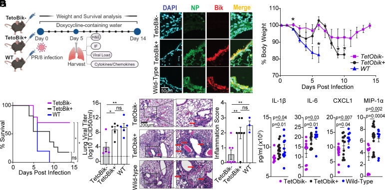
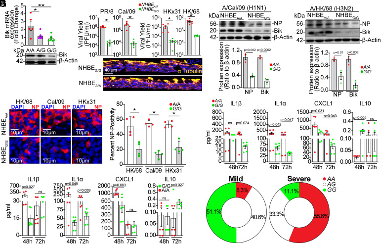
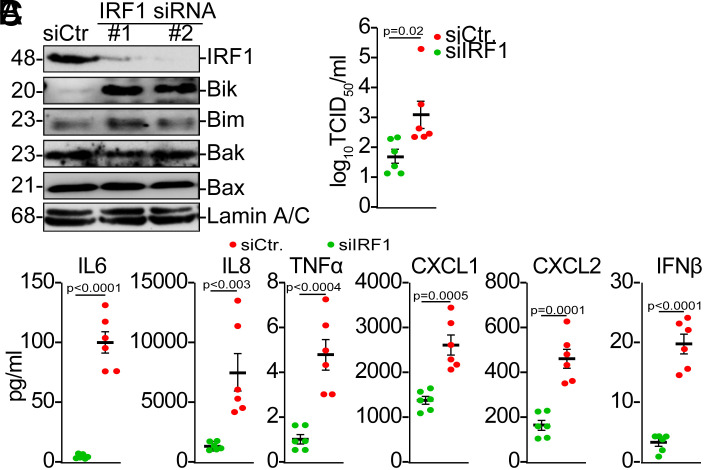
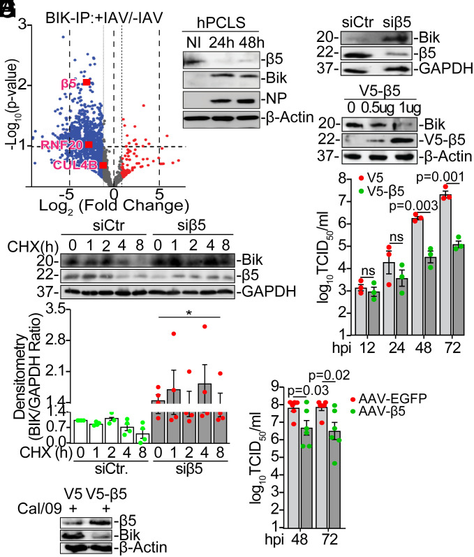
Influenza A viruses (IAVs) pose a significant public health threat, with host factors playing a crucial role in disease severity. We investigated the role of Bcl-2-interacting killer (BIK) in IAV infection using cellular and mouse models, and influenza-infected human cohort. In airway epithelial cells (AECs), BIK deficiency impaired viral replication, while BIK restoration enhanced it. Conversely, airway-specific BIK overexpression in mice increased viral load, inflammation, and mortality, whereas BIK suppression conferred protection. Critically, a genetic variation (rs738276) in the BIK gene, influencing BIK expression, correlates with altered viral replication in air-liquid interface differentiated primary normal human bronchial epithelial cells and influenza severity in humans. Mechanistically, we demonstrate that IAV nucleoprotein (NP) suppresses β5, a subunit of the proteasome, leading to increased BIK levels and enhanced viral replication. Conversely, β5 treatment dampened BIK levels and protected mice from IAV-induced morbidity and mortality. Furthermore, BIK interacts with NP, disrupting the Bcl-2/NP interaction and promoting viral replication. Our findings uncover an IAV-BIK-β5 axis that governs viral replication, suggesting that targeting BIK or β5 may offer therapeutic strategies against influenza.

Keywords: BIK; Beta 5; Influenza A virus; Proteasome; SNP.


Conflict of interest statement

Competing interests statement:The A.G.-S. laboratory has received research support from GSK, Pfizer, Senhwa Biosciences, Kenall Manufacturing, Blade Therapeutics, Avimex, Johnson & Johnson, Dynavax, 7Hills Pharma, Pharmamar, ImmunityBio, Accurius, Nanocomposix, Hexamer, N-fold LLC, Model Medicines, Atea Pharma, Applied Biological Laboratories and Merck, outside of the reported work. A.G.-S. has consulting agreements for the following companies involving cash and/or stock: Castlevax, Amovir, Vivaldi Biosciences, Contrafect, 7Hills Pharma, Avimex, Pagoda, Accurius, Esperovax, Applied Biological Laboratories, Pharmamar, CureLab Oncology, CureLab Veterinary, Synairgen, Paratus, Pfizer and Prosetta, outside of the reported work. A.G.-S. has been an invited speaker in meeting events organized by Seqirus, Janssen, Abbott, Astrazeneca and Novavax. A.G.-S. is inventor on patents and patent applications on the use of antivirals and vaccines for the treatment and prevention of virus infections and cancer, owned by the Icahn School of Medicine at Mount Sinai, New York, outside of the reported work. Y.T. has received research funds from Bayer Pharmaceuticals, AstraZeneca, and Verra Therapeutics. P.G.T. is on the SAB of Immunoscape and Shennon Bio, has received research support and personal fees from Elevate Bio, and consulted for or had travel expenses paid by 10x Genomics, Illumina, Pfizer, Cytoagents, Merck, Bluebird, and JNJ. S.S., S.Y., E.K.A., H.P., M.P., S.E.Z., L.R., V.A., L.A., R.S.N., A.L.M., J.C.H, M.R., Y.T., and Y.A.M. have declared that no conflicts of interest exist. R.A.M. served as a scientific advisor for Valneva SE.

Figures

Fig. 1.
Fig. 1.
BIK is essential for IAVs replication. (A) Cells from ALI-differentiated uninfected WT (bik+/+), bik−/−, and noxa−/− primary MAECs, shown by immunofluorescence (Left panel) were subjected to Western blot (Right panel). α-tubulin was used to stain differentiated cells. (B) ALI-differentiated primary MAECs were infected with 0.1 MOI PR/8 for the indicated time points. Viral titers were analyzed in the apical washes using plaque assay. n = 6/group. One-way ANOVA. (C) ALI-differentiated primary bik+/+ and bik−/− MAECs were infected with 0.1 MOI of the indicated IAVs. Viral titers were analyzed 72 hpi by plaque assay. n = 6/group. Two-tailed t test. (D) Human AECs stably expressing shCtr or shBIK, or bik+/+ and bik−/− MAECs were infected with (E) 1 MOI PR/8 or (F) 1 MOI HK/68 or Cal/09. Protein lysates were analyzed 24 hpi. (G and H) Primary bik−/− MAECs differentiated in ALI were infected with 0.1 MOI PR/8 and treated with 50 MOI of an empty adenoviral vector (Ad-) or Ad-BIK: (G) Protein lysates were analyzed 72 hpi. (H) Viral titers were analyzed in the apical washes 72 hpi by TCID50. n = 6/group. Two-tailed t test. (I) HEK293T cells were infected with lentiviral vector for BIK CRISPR and Cas9. BIK knockout clones were identified through selection using puromycin and BIK expression level was compared between clones by Western blot. Clone #5 was used for the subsequent studies. (J) Parent and CRISPR/Cas9 BIK knockout HEK293T cells were cotransfected with PA, PB1, PB2, and NP plasmids from PR/8 on a pCAGGS backbone, and firefly and renilla luciferase plasmids. The firefly luciferase/renilla ratio is used to calculate the luciferase (polymerase) activity. n = 3/group. Two-tailed t test. Error bars: Mean ± SEM. **P < 0.01; ***P < 0.001. Western blotting experiments were performed in triplicate, and representative images are presented.
Fig. 2.
Fig. 2.
Airway-specific BIK overexpression enhances IAV-induced morbidity and mortality in mice. (A) Eight weeks old WT, CCSP-IndBik transgenic mice in bik−/− background and their littermates were infected with 250 pfu PR/8 intranasally and kept on 400 mg/L doxycycline containing water ad libitum. Figure made with Biorender.com. (B) BIK and viral NP protein expression levels were analyzed in the lungs of mice by immunostaining on 5 dpi. Mice were monitored for (C) changes in body weight (one-way ANOVA) and (D) survival (Log-rank (Mantel-Cox) test) for a period of 14 d. n = 10/group (5 males and 5 females). (E) Lung viral load was analyzed using the median tissue culture infectious dose (TCID50) 5 dpi. The lowest detection limit for the TCID50 is 102. n = 5/group; One-way ANOVA. (F) Microscopic evaluation of lung sections stained with H&E for histopathological analysis at 5 dpi. Red arrows indicate inflammatory cell infiltration in the alveoli and septa. Inflammation score was calculated in a blind manner by a certified pathologist. One-way ANOVA; n = 3 to 7/group. (G) Proinflammatory cytokine and chemokine levels in the lung homogenates analyzed at 5 dpi using Luminex multiplex assay. ANOVA; n = 8 to 16/group. Error bars: Mean ± SEM; *P < 0.05, **P < 0.01, ns = not significant.
Fig. 3.
Fig. 3.
BIK SNP affects its expression and IAV replication. Primary NHBEs from people with AA, AG, or GG variant of BIK SNP (rs738276) were differentiated in ALI culture. (A) BIK mRNA and protein expression levels were analyzed by qRT-PCR and Western blot, respectively. (B) Differentiated primary NHBEs cultures from individuals with AA or GG variants of the BIK SNP were infected with 0.1 MOI PR/8, Cal/09, HKx31, or HK/68. Apical washes were analyzed for virus titers using plaque assay 72 hpi. The tissue section below the graph shows staining of the differentiated cultures for α-tubulin (marker for ciliated cells) and DAPI (nuclear staining). Two-tailed t test; n = 6/group. (C and D) Protein lysates from differentiated cultures infected with (C) H1N1 or (D) H3N2 subtypes were analyzed for viral NP and BIK levels 24 hpi. Relative protein expression was calculated by densitometric analysis (ratio to β-actin) and normalized to AA. n = 3/group. (E) Differentiated cultures from the indicated BIK SNP were infected with the indicated strains of IAV and immunostained for viral NP and analyzed by confocal microscopy 24 h later. Percent NP-positivity was analyzed using ImageJ software. Two-tailed t test; n = 4/group. (F and G) Inflammatory cytokine and chemokine levels in the supernatants of NHBEs infected with (F) Cal/09 or (G) HK/68 were analyzed using Luminex multiplex assay. t test; n = 6/group. (H) Genotype frequency distribution of rs738276 in FLU09 cohort of naturally acquired influenza infection (n = 105) in mild influenza illness phenotypes (Left) compared with severe influenza illness phenotypes (Right). Western blotting experiments were performed in triplicate, and representative images are presented. Error bars: Mean ± SEM. *P < 0.05, **P < 0.01.
Fig. 4.
Fig. 4.
IRF1 silencing upregulates BIK, promoting viral replication and inflammation. NHBEs with the GG variant of the BIK SNP (NHBEG/G) were transfected with siControl or siIRF1 followed by infection with 0.1 MOI Cal/09 48 h posttransfection. (A) Protein lysates were analyzed for BIK, Bim, Bax, Bak, and IRF1 levels. (B) Viral titers and (C) inflammatory cytokine and chemokine levels in cell supernatants were compared at 72 hpi using TCID50 assay and Luminex multiplex assay, respectively. t test; n = 6/group. Error bars: Mean ± SEM.
Fig. 5.
Fig. 5.
IAV-NP inhibits proteasomal degradation of BIK. (A) HAECs were infected with 0 to 2 MOI Cal/09 and protein lysates analyzed for BIK, NP, and β5 expression. (B) HEK293T cells were transfected with 1 µg EV or the indicated viral protein constructs from Cal/09. (C) HEK293T cells were transfected with 0-2 µg Cal/09 NP plasmid. Protein lysates were analyzed for BIK, β5, and viral protein expression levels. (D) HEK293T cells were treated with vehicle or 20 µM MG132 for 6 h. Protein lysates were analyzed for BIK expression level. (E and F) HEK293T cells were (E) infected with mock (infection media) or 0.1 MOI Cal/09 or (F) transfected with 1.0 µg EV or Cal/09 NP-expressing plasmid. Forty-eight hours later, the cells were treated with 20 µM MG132 for 6 h. Protein lysates were immunoprecipitated with anti-BIK antibody and analyzed for BIK ubiquitination using anti-ubiquitin antibody. (G and H) HEK293T cells were (G) infected with mock or 0.1 MOI Cal/09 or (H) transfected with EV or Cal/09 NP-expressing plasmid. Forty-eight hours later, protein lysates were immunoprecipitated with (G) anti-BIK or (H) anti-K48 ubiquitin antibodies and were analyzed for (G) K48 or (H) BIK. Western blotting experiments were performed in triplicate, and representative images are presented.
Fig. 6.
Fig. 6.
IAV-NP inhibits β5 to stabilize BIK and promote viral replication. (A) HEK293T cells were infected with vehicle or 1 MOI Cal/09. Protein lysates were immunoprecipitated with anti-BIK antibody and resolved using SDS-PAGE. An equal amount of BIK was pulled from vehicle- or Cal/09-infected cells and submitted for proteomics analysis. Protein bands were cut and subjected to Mass Spectrometry analysis. The Volcano plot was generated using GraphPad Prism software to identify the top BIK-interacting proteins increased or inhibited by IAV infection. n = 3/group. (B) Human precision-cut lung slices (hPCLS) were infected with mock (PBS, no infection (NI)) or Cal/09 for 24 and 48 h. Protein lysates were analyzed for the expression of β5, BIK, and NP. (C) NHBEs were transfected with siControl or siβ5. Forty-eight hours later protein lysates were analyzed for BIK and β5 levels. (D) NHBEs were transfected with 0 to 1 µg V5-β5. Forty-eight hours later protein lysates were analyzed for BIK and β5 levels. (E) A549 cells were transfected with siCtr or siβ5. Forty-eight hours later, cells were treated with 50 µg/mL cycloheximide (CHX) for the indicated time in hours (h). Protein lysates were analyzed for the expression levels of BIK and the half-life of BIK was compared. *P < 0.05. (F and G) A549 cells were transfected with V5 or V5-β5 followed by infection with 0.1 MOI Cal/09. (F) Viral titers in the apical washes were compared at the indicated time points using TCID50 assay. (G) Cell lysates were analyzed for BIK and β5 expression at 72 hpi. Two-tailed t test. (H) ALI-differentiated NHBEA/A cultures were infected with 0.1 MOI Cal/09 and treated with 1011 genome copies/well of AAV-GFP or AAV-β5. Viral titers in the apical washes were compared at the indicated time points using TCID50 assay. n = 6/group. Two-tailed t test. Error bars: Mean ± SEM. Western blotting experiments were performed in triplicate, and representative images are presented.
Fig. 7.
Fig. 7.
β5 protects mice from IAV-induced morbidity and mortality. WT C57BL/6 mice were instilled intranasally with 1011 GC of AAV6.2-EGFP or AAV6.2-β5. Mice were infected with 250 pfu PR/8 on day 5 post-AAV treatment. On day 5 post PR/8 infection, (A) paraffin-embedded lung tissues were immunostained for EGFP or β5 and analyzed using fluorescent microscopy and (B) lung viral load was measured using TCID50 assay. (C and D) Mice were weighed daily and monitored for survival over a period of 14 dpi. n = 10 to 13/group, *P < 0.05. (E) Inflammatory cytokine and chemokine levels in the lung homogenates analyzed at 5 dpi using Luminex multiplex assay. t test; n = 4-10/group. Error bars: Mean ± SEM.
Fig. 8.
Fig. 8.
BIK/NP interaction stabilizes vRNP. (A) HAECs were infected with the vehicle or 0.1 MOI PR/8 and protein lysates were treated with RNase A. The NP and BIK immunoprecipitates (IPs) were analyzed for BIK and NP levels. (B) bik+/+ and bik−/− MAECs were infected with 1 MOI PR/8. Protein lysates were treated with RNase A. NP and Bcl-2 IPs were analyzed. (C) HAECs were infected with 0.1 MOI PR/8 and 24 h later treated with empty adenoviral vector (Ad-) or 50 MOI Ad-BIK. Protein lysates were treated with RNase A. Bcl-2 IP was analyzed for NP and BIK protein levels. (D) HAECs were infected with 0.1 MOI PR/8 and 24 h later with 50 MOI Ad-BIKL61G (with impaired interaction with Bcl-2) or Ad-BIKWT. Protein lysates were treated with RNase A. Bcl-2 IPs were analyzed for NP and BIK protein levels. (E) HEK293T cells were transfected with EV or Bcl-2 expression vector and infected with 0.1 MOI PR/8 24 h later. Protein lysates were analyzed for the level of NP and Bcl-2 24 hpi. (F) HEK293T cells transfected with EV or Bcl-2 expression vector and infected with 0.1 MOI of the indicated IAV strains. Virus titers were compared in the apical washes using plaque assay at 72 hpi. *P < 0.05. (G and H) HEK293T cells were transfected with 1 µg EV, WT-, Mut1 (E46A, K48A)-, or Mut2 (D101A)-NP expressing constructs. (G) Cell lysates were analyzed for BIK and NP levels. (H) Protein lysates were immunoprecipitated with anti-NP or anti-BIK antibodies and analyzed for BIK/NP interaction. Western blotting experiments were performed in triplicate, and representative images are presented. Error bars: Mean ± SEM.

References

    1. Lampejo T., Influenza and antiviral resistance: An overview. Eur. J. Clin. Microbiol. Infect. Dis. 39, 1201–1208 (2020). - PMC - PubMed
    1. Fiore A. E., et al. , Prevention and control of influenza: Recommendations of the Advisory Committee on Immunization Practices (ACIP), 2008. MMWR Recomm. Rep. 57, 1–60 (2008). - PubMed
    1. Molinari N. A., et al. , The annual impact of seasonal influenza in the US: Measuring disease burden and costs. Vaccine 25, 5086–5096 (2007). - PubMed
    1. Hayden F. G., Perspectives on antiviral use during pandemic influenza. Philos. Trans. R. Soc. Lond. B, Biol. Sci. 356, 1877–1884 (2001). - PMC - PubMed
    1. Davies W. L., et al. , Antiviral activity of 1-adamantanamine (amantadine). Science 144, 862–863 (1964). - PubMed

MeSH terms