. 2025 Jul 8;15(1):24411.
doi: 10.1038/s41598-025-02010-1.

CLN3 disease disrupts very early postnatal hippocampal maturation

Affiliations

CLN3 disease disrupts very early postnatal hippocampal maturation

Jeet B Singh et al. Sci Rep. .

Erratum in

Abstract

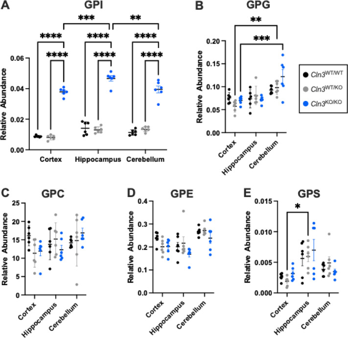
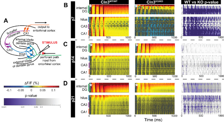
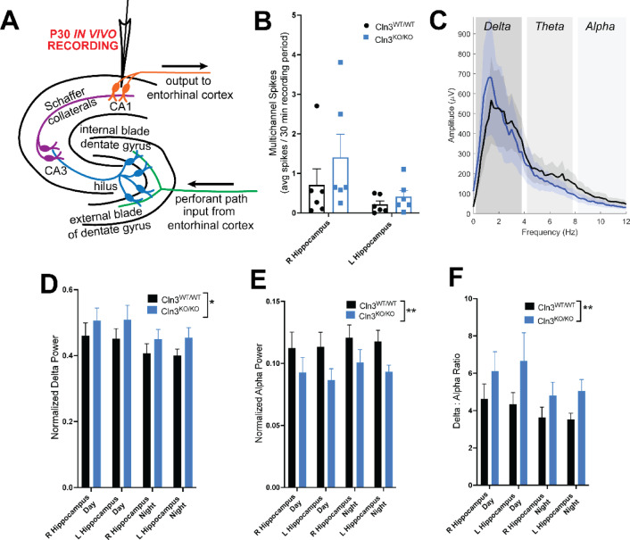
CLN3 disease or juvenile neuronal ceroid lipofuscinosis (Batten disease), is a progressive, severe, neurodegenerative, lysosomal storage disorder. Previous studies have demonstrated that network-level excitability differences are present in mouse models prior to significant lysosomal storage accumulation. Here we sought to identify the earliest biochemical and functional markers of disease in the hippocampus, a brain region important in learning and memory and implicated in CLN3 disease. Using targeted hydrophilic interaction liquid chromatography high resolution mass spectrometry (LC-HRMS), we quantified levels of glycerophosphodiesters (GPDs), recently-described biomarkers of CLN3 disease, in early postnatal hippocampus. In addition, we assessed hippocampal excitability via in vitro voltage-sensitive dye imaging (VSDI) across the period of postanal hippocampal maturation (p7, p14, p21). Finally, we completed longitudinal electroencephalogram (EEG) recordings to evaluate in vivo hippocampal circuit dynamics once the hippocampal circuit was matured. Intriguingly, glycercophosphoinositol (GPI or GroPIns), but not other GPDs, were significantly elevated in CLN3 disease hippocampus in early development at p11, further supporting the hypothesis that GPI plays a key role in disease pathogenesis. Functionally, the hippocampus was significantly hypoexcitable as early as p7 and showed a very atypical pattern of maturation across early development. This aberrant development resulted in abnormal in vivo circuit function, with pathologic slowing observed on EEG recordings at p30. Collectively these data underscore the potential link between pathologic metabolism of GPI and functional defects in CLN3 disease. In addition, this work highlights that CLN3 disease is an early neurodevelopmental, and not just neurodegenerative, disorder.

Keywords: CLN3 disease; Glycerophosphodiesters (GPDs); Lysosomal storage disorders; Neurodevelopmental disorders.


Conflict of interest statement

Declarations. Competing interests: The authors declare no competing interests. Ethical approval: All procedures performed in studies involving animals were in accordance with the CHOP Institutional Animal Care and Use Committee.

Figures

Fig. 1
Fig. 1
CLN3 disease mice accumulate GPIs, but not other GPDs, by p11 throughout the brain. HILIC method with full scan in negative mode was performed on individual brain regions from Cln3−/− (blue), Cln3−/+ (gray), and Cln3−/+ (black) mice. (A) Gycerophosphoinositol (GPI) (B) glycerophosphoglycerol (GPG), (C) Glycerophosphocholine (GPC), (D) glycerophosphoethanolamine (GPE), and Glycerophosphoserine (GPS). Data are mean ± s.e.m. (n = 6 mice per genotype; 3 female, 3 male). 2-Way ANOVA with multiple comparisons was performed between all genotypes within a brain region and between the same genotypes between different brain regions (p values; *< 0.05, ** <0.01, *** < 0.0005, **** <0.0001).
Fig. 2
Fig. 2
CLN3 disease hippocampus is significantly hypoexcitable in response to perforant path (PP) stimulation by p7. (A) The PP was stimulated, and the response was measured using voltage sensitive dye imaging in hippocampal slices from (B) p7, (C) p14, and (D) p21 Cln3−/− and Cln3+/+ mice. Raster plots of average fluorescence change (DF/F, warm colors excitation, cool colors inhibition) over time (x axis) and location within hippocampus (y axis) are shown. Stimulation of Cln3+/+ slices results in robust excitation of the dentate gyrus (DG) that spreads to the CA proper (hilus, CA3, CA1) at p7. In Cln3+/+ slices from older (p14 and p21) animals demonstrates increased gating function of the DG, as the spread of excitation from the DG to the CA proper is reduced. In Cln3−/− animals, the DG is significantly hypoexcitable at p7. DG excitability improves by p14, but by p21 the CA proper region becomes hypoexcitable, consistent with prior studies of older Cln3−/− mice. For each age group, Cln3−/− vs. Cln3+/+ rasters were compared using a permutation sampling method with 1000 iterations. Regions of significant (p < = 0.05) hypoexcitability are shown in purple. N = at least 10 slices from 5 animals per condition.
Fig. 3
Fig. 3
CLN3 disease hippocampus is initially hyperexcitable then hypoexcitable to Schaffer collateral (SC) stimulation. (A) The VSDI response after SC stimulation of (B) p7, (C) p14, and (D) p21 Cln3−/− and Cln3+/+ hippocampus is shown. Raster plots of average fluorescence change (DF/F, warm colors excitation, cool colors inhibition) over time (x axis) and location within hippocampus (y axis) are shown. Stimulation results in both forward and backward propagation of excitation. At p7 and p14 there is a trend toward hyperexcitability in Cln3−/− hippocampus, but by p21, both forward and backward propagation of excitation is decreased. For each age group, Cln3−/− vs. Cln3+/+ rasters were compared using a permutation sampling method with 1000 iterations. Regions of significant (p < 0.05) hypoexcitability are shown in purple. N = at least 10 slices from 5 animals per condition. Location of stimulus shown by an asterix.
Fig. 4
Fig. 4
p30 Cln3 disease hippocampus shows background slowing with an increased delta / alpha ratio on EEG. (A) Long-term in vivo EEG recordings from the CA1 region of the hippocampus from p30 mice shows no significant difference in (B) interictal spikes or (C) total raw power between Cln3+/+ (black) and Cln3−/− (blue) hippocampus. However, there is significant background slowing in Cln3−/− hippocampus as indicated by a (D) increase in slow delta band (0.1–4 Hz) power, a (E) decrease in alpha band (8–12 Hz) power, and a resulting (F) increased delta: alpha ratio. Data were analyzed via 2-way ANOVA, with p-value for genotype as a source of variation shown, *p < = 0.05, **p < = 0.01. N = 7 Cln3+/+ and 6 Cln3−/− mice. Spikes were quantified as spikes / 30 min period averaged from 24 h of recording. Average power in each frequency band was calculated for 12 h of daytime and 12 h of night recordings from electrodes implanted into the right and left hippocampi.

References

    1. Laqtom, N. N. et al. CLN3 is required for the clearance of glycerophosphodiesters from lysosomes. Nature609(7929), 1005–1011 (2022). - PMC - PubMed
    1. Heins-Marroquin, U. et al. CLN3 deficiency leads to neurological and metabolic perturbations during early development. Life Sci. Alliance7(3) (2024). - PMC - PubMed
    1. Brudvig, J. J. et al. Glycerophosphoinositol is elevated in blood samples from CLN3 (Deltaex7-8) pigs, Cln3 (Deltaex7-8) mice, and CLN3-affected individuals. Biomark. Insights17, p11772719221107765 (2022). - PMC - PubMed
    1. Carcel-Trullols, J., Kovacs, A. D. & Pearce, D. A. Cell biology of the NCL proteins: What they do and don’t do. Biochim. Biophys. Acta1852(10 Pt B), 2242–2255 (2015). - PubMed
    1. Ku, C. A. et al. Detailed clinical phenotype and molecular genetic findings in CLN3-associated isolated retinal degeneration. JAMA Ophthalmol.135(7), 749–760 (2017). - PMC - PubMed

MeSH terms

LinkOut - more resources