Review
. 2025 Jul;57(7):1467-1481.
doi: 10.1038/s12276-025-01497-y. Epub 2025 Jul 8.

Structural insights into GPCR signaling activated by peptide ligands: from molecular mechanism to therapeutic application

Affiliations
Review

Structural insights into GPCR signaling activated by peptide ligands: from molecular mechanism to therapeutic application

Jinuk Kim et al. Exp Mol Med. 2025 Jul.

Abstract

Recent advances in structural biology have profoundly enhanced our understanding of G protein-coupled receptors (GPCRs), providing detailed molecular insights into their activation and ligand recognition. Here, in this Review, we explore the molecular mechanisms of class A and class B GPCRs bound to peptide agonists and their implications for drug development. We examine representative GPCRs, such as the angiotensin II type 1 receptor, chemokine receptor 5, μ-opioid receptor, parathyroid hormone 1 receptor and glucagon-like peptide 1 receptor (GLP-1R), highlighting their activation processes upon peptide ligand binding. Comparative analysis of structures bound to endogenous and synthetic peptide ligands reveals critical insights for rational drug design. A case study on GLP-1R demonstrates how structural insights have led to the design of successful drugs for type 2 diabetes and obesity. This comparative structural analysis aims to deepen our understanding of GPCR activation mechanisms and support future drug discovery efforts targeting peptide-binding GPCRs.


Conflict of interest statement

Competing interests: The authors declare no competing interests.

Figures

Fig. 1
Fig. 1. Diverse physiological roles of GPCR-targeting peptides around the human body.
Representative GPCR-targeting peptides and their associated physiological roles are summarized across various regions of the human body. The structure of each peptide represents its conformation when bound to its respective GPCR (PDB: (brain) 7W53, 7L1U, 8JBG, 8GY7, 8F7Q, 7F9Y, 7XJJ and 7VGX; (heart) 6JOD, 8XZG and 6PD1; (spinal cord) 8H0P; (kidney) 7DW9, 7TYO and 8FLU; (gut) 7EZH and 8IBV; (liver) 6LML; (uterus) 8I2G and 7RYC; (immune) 8HK5, 7SK3 and 7YKD, from left to right). Peptides are shown as cartoons or stick representations.
Fig. 2
Fig. 2. Diverse binding modes of peptide ligands to their GPCRs.
Structures of five representative GPCRs in their peptide ligand-bound states are presented: AT1R (6OS0), CCR5 (7O7F), MOR (8F7R), PTH1R (7VVL), and GCGR (6LMK). Receptors are shown as cartoons in distinct colors, and G proteins (or the nanobody in PDB 6OS0) are omitted for clarity. Peptide ligands, angiotensin II (AT2), CCL5, endomorphin, PTH and glucagon, are displayed as surface models.
Fig. 3
Fig. 3. General activation mechanisms of class A and B GPCRs bound to peptide ligands.
a A schematic representation of the inactive and active states of class A GPCRs. While small-molecule antagonists typically bind into the transmembrane (TM) pocket stabilizing the receptor in an inactive state, peptide agonists, generally, interact with ECL2 and the N-tail, which are flexible in the absence of a peptide ligand (indicated by a dashed black arrow), as well as with the TM pocket. Agonist binding induces conformational changes in the TMD, resulting in the outward movement of the cytoplasmic segments of TM5 and TM6 (indicated by a red arrow), thereby facilitating G protein binding. b A schematic representation of the inactive and active states of class B GPCRs. These receptors possess a large N-terminal ECD, which is flexible in the absence of a peptide ligand (dashed black arrow). Peptide agonists engage both the ECD and TMD. In the active state, a sharp kink or bend in the PxxG motif of TM6 promotes the outward movement of its cytoplasmic segment (red arrow), creating an open cavity for G protein binding.
Fig. 4
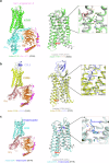
Fig. 4. Activation mechanisms of peptide-binding class A GPCRs.
a Left: active structure of the Sar1-angiotensin II‒AT1R‒Gq complex (PDB: 7F6G), color-coded as follows: Sar1-Angiotensin II (pink), AT1R (green), Gαq (cyan), Gβ (orange) and Gγ (magenta). Middle: structural comparison of the active (green, PDB: 6OS0) and inactive (gray, PDB: 4YAY) states of AT1R. Right: close-up view of the binding pocket highlighting the critical role of the C-terminal phenylalanine (F8) of angiotensin II in receptor activation. Conformation changes in key residues are indicated by green arrows, while the red arrow marks the outward movement of TM6 upon activation. b Left: active structure of CCR5 (yellow), in complex with CCL5 (slate) and the Gi heterotrimer (Gαi (salmon), Gβ (orange), and Gγ (magenta)) (PDB: 7O7F). Middle: structural comparison between the active (yellow, PDB: 7O7F) and inactive (gray, PDB: 6AKY) states of CCR5. Right: close-up of the orthosteric pocket showing how the N-terminal region of CCL5 interacts with the receptor. Upon activation, conformational changes in W2486.48 and the outward movement of TM6 are indicated by yellow and red arrows, respectively. c Left: active structure of MOR (cyan) in complex with endomorphin (blue) and the Gi heterotrimer (PDB: 8F7R). Middle: structural comparison between the active (cyan, PDB: 8F7R) and inactive (gray, PDB: 4DKL) states of MOR. Right: close-up of the orthosteric pocket showing the binding of Y1 of endomorphin (blue). The red arrow highlights the outward movement of TM6 during activation.
Fig. 5
Fig. 5. Structural comparison of endogenous and synthetic ligands bound to class A GPCRs.
a Superposition of MIP-1α-bound active and maraviroc-bound inactive structures of CCR5. The receptor is shown as a cartoon with α-helices depicted as cylindrical helices. Ligands are shown as sticks and color-coded as follows: active CCR5 (pale cyan), MIP-1α (slate), inactive CCR5 (light orange) and maraviroc (orange). A close-up view of the TM pocket is shown on the right. The N-terminal region of MIP-1α binds deeply into the TM pocket, with maraviroc occupying a similar position. b Superposition of GP120-bound and maraviroc-bound inactive structures of CCR5. The receptor is shown as a cartoon and ligands are represented as sticks. GP120-bound CCR5 and GP120 are colored light red and cyan, respectively. For clarity, CD4 has been omitted. Maraviroc-bound CCR5 and maraviroc are colored light orange and orange, respectively. The binding pocket of CCR5 is highlighted in a zoomed-in view. c Superposition of endomorphin-bound and fentanyl-bound active structures of MOR. The receptor is shown as a cartoon, and ligands are shown as sticks, color-coded as follows: endomorphin-bound MOR (pale green), endomorphin (green), fentanyl-bound MOR (light pink) and fentanyl (yellow). A close-up of the TM pocket highlights the binding positions of the two MOR ligands.
Fig. 6
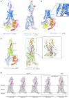
Fig. 6. Activation and therapeutic targeting of peptide-binding class B GPCRs.
a The active structure of the PTH‒PTH1R‒Gs complex is presented, with components color-coded as follows: PTH (violet), PTH1R (blue), Gαs (blue white), Gβ (orange), Gγ (magenta) and Nb35 (yellow). A structural comparison of the preactive and active states of PTH1R is shown on the right. The G protein is omitted for clarity. The conformational changes from preactive to active states are indicated with red arrows. b The active structure of PTH1R bound to PCO371 is shown. PCO371 binds to an intracellular allosteric site, distinct from the orthosteric peptide-binding pocket. PCO371 is shown as salmon-colored sticks. A zoomed-in view on the right highlights the novel binding site of PCO371. c The active structure of GLP-1R (lime) bond to GLP-1 (warm pink) is shown on the left. Superposition of the active and inactive GLP-1R structures is shown on the right (G protein omitted for clarity). ECD, which undergoes dramatic rotation upon GLP-1 binding, is shown in orange (active) and pale cyan (inactive), and the movements of the ECD and TM6 upon activation are indicated with green and red arrows, respectively. d Structures of GLP-1R bound to endogenous ligand GLP-1 and various agonist drugs are presented. A comparison of GLP-1R and GIPR structures, both bound to the dual-agonist tirzepatide, is shown on the right. Receptors are displayed as transparent surfaces with light-gray-colored cartoons, and ligands are shown as magenta cartoons.

Similar articles

References

    1. Zhang, M. Y., Chen, T., Lu, X., Lan, X. B., Chen, Z. Q. & Lu, S. Y. G protein-coupled receptors (GPCRs): advances in structures, mechanisms, and drug discovery. Signal Transduct. Target. Ther.9, 88 (2024) - PMC - PubMed
    1. Stevens, R. C. et al. The GPCR Network: a large-scale collaboration to determine human GPCR structure and function. Nat. Rev. Drug Discov.12, 25–34 (2013). - PMC - PubMed
    1. Sutkeviciute, I. & Vilardaga, J. P. Structural insights into emergent signaling modes of G protein-coupled receptors. J. Biol. Chem.295, 11626–11642 (2020). - PMC - PubMed
    1. DeMaria, S. & Ngai, J. The cell biology of smell. J. Cell Biol.191, 443–452 (2010). - PMC - PubMed
    1. Roper, S. D. & Chaudhari, N. Taste buds: cells, signals and synapses. Nat. Rev. Neurosci.18, 485–497 (2017). - PMC - PubMed

LinkOut - more resources