. 2025 Jul 8;8(1):1017.
doi: 10.1038/s42003-025-08460-7.

Lactoferrin-osteopontin complexes: insights into intestinal organoid bioavailability and gut microbiota modulation

Affiliations

Lactoferrin-osteopontin complexes: insights into intestinal organoid bioavailability and gut microbiota modulation

Camilla Ceroni et al. Commun Biol. .

Abstract

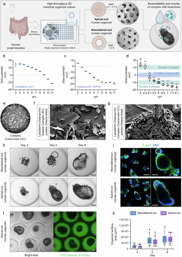
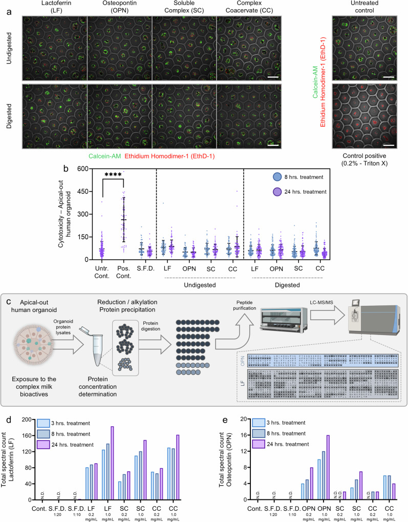
Lactoferrin (LF) and osteopontin (OPN) are key bioactive milk proteins with significant immune-modulatory, gut, and systemic health benefits. We investigated the bioavailability and intestinal uptake dynamics of bovine LF-OPN soluble complexes (SC) and complex coacervates (CC) using a microarrayed high-throughput 3D apical-out intestinal organoid platform, which closely mimics the human intestinal epithelium. Our findings revealed that both SC and CC complexes exhibited cellular uptake compared to individual LF and OPN components. Nevertheless, complexation did not compromise intestinal organoid viability, even following extended exposure. Further, an ex vivo bioreactor-based colon fermentation study using infant fecal microbiota demonstrated that LF-OPN complexes significantly influenced microbial metabolic activities. This modulation leads to enhanced production of short-chain fatty acids (SCFAs), particularly elevating butyrate levels, a key metabolite for sustaining gut health. The phylogenetic analysis highlighted significant shifts in microbial composition, favoring beneficial bacterial families such as Bacteroides fragilis, Phocaeicola dorei, Parabacteroides spp., and Clostridium symbiosum for complex bioactives. Our findings indicate that LF-OPN complexes have significant potential to further optimize infant nutrition by enhancing the bioavailability of bioactive compounds and promoting gut health through microbiota modulation.


Conflict of interest statement

Competing interests: L.D., M.E., M.R-C.K., J.O’R., D.G., and O.M. are or were employees of the Société des Produits Nestlé S.A., Switzerland. C.C., J.D., P.S., A.R., and N.B. are or were employees of DOPPL S.A., Switzerland. P.V.d.A. and A.B. are employees of Cryptobiotix S.A., Belgium.

Figures

Fig. 1
Fig. 1. Development and characterization of a microarrayed high-throughput 3D intestinal organoid culture platform to assess the bioavailability of complex milk bioactives.
a Schematic representation of the high-throughput 3D intestinal organoid culture platform. Human small intestine biopsies are processed to derive apical-out and basolateral-out human intestinal organoids cultured in a microarrayed platform with single-organoid resolution. The application of different concentrations of basement membrane extract (BME) influences the polarity of the organoids, with apical-out organoids being cultured with 0–0.5% BME and basolateral-out organoids with 1.5% BME. This platform enables the evaluation of the bioavailability and toxicity of complex milk bioactives, such as lactoferrin-osteopontin complexes. Created in BioRender. Mashinchian, O. (2025) https://BioRender.com/yyzy0j4. b and c Zeta-potential profiles of bovine lactoferrin (b) and bovine osteopontin (c). Lactoferrin shows a positive zeta potential at lower pH, which decreases as the pH increases, indicating a loss of surface charge. Osteopontin exhibits a similar trend, with a more negative zeta potential as the pH rises. In (b and c), each data point represents the median of an independent experimental run, with each run comprising four technical replicates. For each replicate, 10–100 zeta-potential measurements were recorded per pH value, resulting in a total of ~40–400 data points per pH per independent run. d Zeta-potential of the lactoferrin-osteopontin complex as a function of pH, showing the formation of a soluble complex at higher pH and a transition to a coacervate complex at lower pH (around pH 5–5.8). The shaded regions indicate the pH ranges for the formation of soluble complexes and complex coacervates. Box-and-whisker plots represent the distribution of zeta-potential values from 10–48 independent experimental runs (number of runs per pH indicated above each box), with each run comprising four technical replicates. For each replicate, 10–100 zeta-potential measurements were recorded per pH value. Horizontal lines represent the median, and whiskers extend to the 10 and 90th percentiles. e–g Morphological characterization of lactoferrin-osteopontin complex coacervates. e. Light microscopy image of the lactoferrin-osteopontin complex coacervate (CC) showing spherical structures (40x magnification). f Scanning electron microscopy (SEM) image of freeze-dried lactoferrin-osteopontin soluble complexes (SC) showing a flaky structure. Scale bar = 50 µm. g SEM image of freeze-dried lactoferrin-osteopontin complex coacervates (CC) revealing a highly structured, porous morphology. Scale bar = 50 µm. h Bright-field images of basolateral-out and apical-out human intestinal organoids over eight days. Scale bar = 100 µm. i Immunofluorescent staining of basolateral-out and apical-out human intestinal organoids for F-actin (green) and DNA (blue), highlighting structural differences in polarity and surface area between the two organoid types. Scale bar (left overview): 200 µm; Scale bar (right, single organoid): 100 µm. j Permeability assay of apical-out human intestinal organoids using FITC-Dextran (4.4 kDa). Bright-field and fluorescence images show the diffusion of FITC-Dextran within the organoids. Scale bar = 100 µm. k Box-and-whisker plots depict the distribution of organoid surface area (µm²) for basolateral-out (blue; n = 35) and apical-out (purple; n = 41) organoids on days 3, 5, and 8 of culture. Each data point represents an individual organoid measurement. Horizontal lines represent the median and quartiles, and whiskers extend to the 10 and 90th percentiles. Dots represent individual data points beyond this range. Both organoid types show an increased surface area over time, with overlapping size distributions across polarity orientations.
Fig. 2
Fig. 2. Impact of lactoferrin (LF), osteopontin (OPN), and their complexes on apical-out human intestinal organoid viability and uptake using microarrayed high-throughput platform.
a Representative fluorescence microscopy images showing the viability of apical-out human gut organoids following treatment with undigested and digested forms of lactoferrin (LF), osteopontin (OPN), soluble complexes (SC), and complex coacervates (CC). Viability was assessed using Calcein-AM (green), indicating live cells, and Ethidium Homodimer-1 (EthD-1, red), indicating dead or damaged cells. Untreated controls (Untr.) and positive controls (Triton X-treated) are included as references. Scale bar = 500 µm. b Quantitative analysis of the cytotoxicity of apical-out human gut organoids (n = 63 (min) – 164 (max) per condition) treated with LF, OPN, SC, and CC, either undigested or digested, for 8 or 24 h. Organoid cytotoxicity was determined by fluorescence intensity quantification of Calcein-AM and EthD-1 staining. Significant differences between positive control (Triton X-treated) and untreated controls (Untr.) are shown (****p < 0.0001). Two-way ANOVA with Dunnett’s post-test was performed using GraphPad Prism. The graph displays individual organoid data points, mean values, and standard deviation (SD). S.F.D. represents a Sub-free digesta as a control. c Schematic overview of the experimental workflow for assessing the protein uptake of lactoferrin (LF) and osteopontin (OPN) by human intestinal organoids. Apical-out human organoids were exposed to the bioactive milk complexes for 3, 8, and 24 h. Organoid lysates were then processed for proteomic analysis, involving protein reduction/alkylation, precipitation, and digestion with trypsin, followed by peptide purification and LC-MS/MS analysis. Created in BioRender. Mashinchian, O. (2025) https://BioRender.com/yyzy0j4. d Total spectral count of lactoferrin (LF) peptides detected by LC-MS/MS in organoid lysates following 3, 8, and 24 h of treatment with LF, SC, and CC at two concentrations (0.2 and 1.0 mg/mL). S.F.D. controls are shown for comparison. e Total spectral count of osteopontin (OPN) peptides detected by LC-MS/MS in organoid lysates following 3, 8, and 24 h of treatment with OPN, SC, and CC at two concentrations (0.2 and 1.0 mg/mL). S.F.D. controls are shown for comparison.
Fig. 3
Fig. 3. Effects of milk bioactive complexes on microbial activity and composition in an ex vivo infant gut model.
a Overview of the experimental workflow. Fecal samples were collected from infants (2–4 months old; n = 6), and the microbiota was incubated in SIFR® high-throughput bioreactors for ex vivo colon fermentation. Milk bioactive complexes, including lactoferrin (LF), osteopontin (OPN), and their complex formats (SC and CC), were introduced to simulate microbial metabolic interactions and were incubated with the fecal microbiota at 37 °C for 24 h. Post-incubation, microbial metabolite production was analyzed using gas chromatography (GC), and the phylogenetic composition of microbiota was assessed through shallow shotgun sequencing. Created in BioRender. Mashinchian, O. (2025) https://BioRender.com/6i15fh1. b–e Microbial metabolic outcomes from ex vivo colon fermentation across different treatments were measured. Violin plots illustrate physicochemical parameters, including pH (b), gas production (c), branched-chain fatty acids (BCFAs) levels (d), and total short-chain fatty acids (SCFAs) levels (e). Different conditions are shown: control (Cont.), LF, OPN, SC, and CC. Data indicate significant changes in all parameters across different treatments, with specific variations highlighted for each treatment. f–h Violin plots showing the levels of key SCFA: acetate (f), propionate (g), and butyrate (h). All treatments significantly altered SCFA levels compared to the control, with distinct profiles observed for LF and OPN. i Bacterial cell density was measured across different conditions, indicating that the treatments significantly modulated the microbial population, as compared to the control. Significant differences were determined via repeated measures of ANOVA analysis (with Benjamini-Hochberg correction). j Absolute levels (cells/mL) of bacterial phyla in fecal samples after fermentation with different bioactive treatments, including Actinomycetota, Bacteroidota, Bacillota, Pseudomonadota, and Verrucomicrobia. Each treatment altered the microbiota composition. k Principal component analysis (PCA) showing microbiota shifts based on family-level taxonomic data. Distinct clustering of treatment groups was observed, particularly for the LF, OPN, SC, and CC conditions. Key bacterial families contributing to the variance include Bifidobacteriaceae, Lachnospiraceae, Bacteroidaceae, and Rikenellaceae. l Heatmap representing the relative abundance of specific bacterial families within the Bacteroidota and Bacillota phyla across treatments, compared to the control group. The log2-transformed fold changes for each treatment compared to Cont. are color-coded, with green and yellow shades indicating higher abundance compared to controls and blue shades indicating lower abundance. Values in bold highlight significant treatment effects according to repeated measures ANOVA analysis (with Benjamini-Hochberg correction, FDR = 0.20).

Similar articles

References

    1. Lönnerdal, B. Infant formula and infant nutrition: bioactive proteins of human milk and implications for composition of infant formulas. Am. J. Clin. Nutr.99, 712S–717S (2014). - PubMed
    1. Lönnerdal, B. Bioactive proteins in human milk: health, nutrition, and implications for infant formulas. J. Pediatr.173, S4–S9 (2016). - PubMed
    1. Carr, L. E. et al. Role of human milk bioactives on infants’ gut and immune health. Front. Immunol.12, 604080 (2021). - PMC - PubMed
    1. de Weerth, C. et al. Human milk: From complex tailored nutrition to bioactive impact on child cognition and behavior. Crit. Rev. Food Sci. Nutr.63, 7945–7982 (2023). - PubMed
    1. Legrand, D. Overview of lactoferrin as a natural immune modulator. J. Pediatr.173, S10–S15 (2016). - PubMed

LinkOut - more resources