

Validation of noninvasive indices of right ventricular diastolic function. Simultaneous echocardiography and pressure-volume catheterization studies
- PMID: 40629401
- PMCID: PMC12239245
- DOI: 10.1186/s12947-025-00351-5
Validation of noninvasive indices of right ventricular diastolic function. Simultaneous echocardiography and pressure-volume catheterization studies
Abstract
Background: The reliability of the recommended echocardiographic methods for assessing RV diastolic function has been questioned. We aimed to validate noninvasive indices of RV diastolic function, derived from tricuspid Doppler and myocardial deformation metrics, against intrinsic diastolic chamber properties and filling pressures.
Methods: We obtained simultaneous high-fidelity pressure-volume loops and echocardiographic data in separate animal and clinical settings: (1) a porcine model of acute hemodynamic interventions (n = 13), and (2) patients with Fallot tetralogy and pulmonary hypertension (n = 9). These designs allow for within- and between-subject validation. From the PV loops data, we obtained the reference values of RV stiffness (S+), elastic recoil (S-) and relaxation (τ) constants, as well as the contribution of passive properties to instantaneous diastolic pressures.
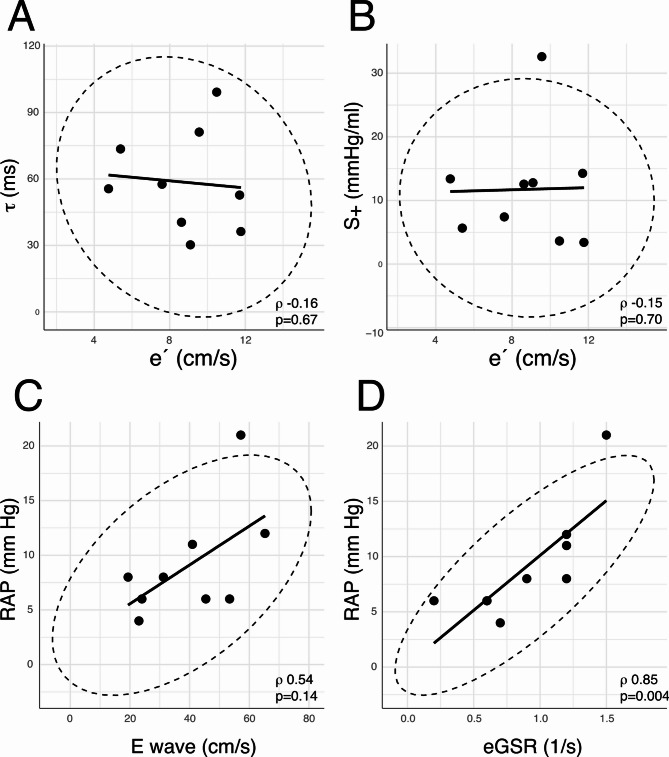
Results: In the animal setting, only the tricuspid E/A ratio and e' velocity weakly correlated with S+ (Rrm:0.36 and 0.28 respectively, p < 0.01 for both). In the clinical group, no correlation was found between the echocardiographic indices and the intrinsic diastolic properties. Isovolumic relaxation time and early diastolic global strain-rate (GSR) correlated with mean right atrial pressure (RAP) (Spearman r: -0.73 and 0.85, respectively, p < 0.05 for both). E/e' and E/GSR ratio were not associated with RAP. Tricuspid e' and GSR negatively correlated with passive pressure component (only due to) at valve opening (Rrm -0.27 and - 0.33, respectively, p < 0.01 for both).
Conclusions: Recommended echocardiographic indices of RV diastolic function do not reflect intrinsic RV diastolic properties. Therefore, the application of these indices for inferring RV diastolic function and filling pressures is limited.
Keywords: Diastolic function; Echocardiography; Elastic recoil; Relaxation; Right ventricle; Stiffness.
© 2025. The Author(s).
Conflict of interest statement
Declarations. Ethics approval and consent to participate: The clinical study protocol complied with the Declaration of Helsinki and was approved by the Committee on Ethics in Drug Research of the Gregorio Marañón Health Research Institute (282/12 and 335/16). All patients provided written informed consent to participate in the study. Competing interests: The authors declare no competing interests.
Figures


Similar articles
-
Right Ventricular Myocardial Global Longitudinal Strain Assessment of Right Ventricular Function in Patients with Pulmonary Embolism.Kardiologiia. 2025 Aug 6;65(7):46-54. doi: 10.18087/cardio.2025.7.n2860. Kardiologiia. 2025. PMID: 40771167
-
Accuracy of Intraoperative Transesophageal Echocardiographic Doppler Parameters in Assessing the Right Ventricular Diastolic Function After Repair of Tetralogy of Fallot in Pediatric Patients.Ann Card Anaesth. 2025 Jan 1;28(1):53-60. doi: 10.4103/aca.aca_85_24. Epub 2025 Jan 24. Ann Card Anaesth. 2025. PMID: 39851150 Free PMC article.
-
Impact of Length Indexing of Deformation in Echocardiographic Evaluation of Right Ventricular Function.J Am Soc Echocardiogr. 2025 Mar;38(3):187-194. doi: 10.1016/j.echo.2024.11.011. Epub 2024 Dec 17. J Am Soc Echocardiogr. 2025. PMID: 39701430
-
What Is the Evidence That the Tissue Doppler Index E/e' Reflects Left Ventricular Filling Pressure Changes After Exercise or Pharmacological Intervention for Evaluating Diastolic Function? A Systematic Review.J Am Heart Assoc. 2017 Mar 15;6(3):e004766. doi: 10.1161/JAHA.116.004766. J Am Heart Assoc. 2017. PMID: 28298372 Free PMC article.
-
Correlation Between Tissue Doppler Imaging Method (E/e') and Invasive Measurements of Left Ventricular Filling Pressures: A Systematic Review, Meta-Analysis, and Meta-Regression.J Cardiothorac Vasc Anesth. 2024 Dec;38(12):3200-3214. doi: 10.1053/j.jvca.2024.08.014. Epub 2024 Aug 13. J Cardiothorac Vasc Anesth. 2024. PMID: 39218765
References
-
- Haddad F, Doyle R, Murphy DJ, Hunt SA. Right ventricular function in cardiovascular disease, part II: pathophysiology, clinical importance, and management of right ventricular failure. Circulation. 2008;117(13):1717–31. - PubMed
-
- Weatherald J, Boucly A, Chemla D, Savale L, Peng M, Jevnikar M, et al. Prognostic value of Follow-Up hemodynamic variables after initial management in pulmonary arterial hypertension. Circulation. 2018;137(7):693–704. - PubMed
-
- Rommel KP, von Roeder M, Oberueck C, Latuscynski K, Besler C, Blazek S, et al. Load-Independent systolic and diastolic right ventricular function in heart failure with preserved ejection fraction as assessed by resting and handgrip exercise Pressure-Volume loops. Circ Heart Fail. 2018;11(2):e004121. - PubMed
Publication types
MeSH terms
Grants and funding
LinkOut - more resources
Full Text Sources
Medical
