. 2025 Jul 8;23(1):758.
doi: 10.1186/s12967-025-06816-5.

CIITA-modified glioblastomas vaccinate and induce cross-protection against heterologous wild-type glioblastomas

Affiliations

CIITA-modified glioblastomas vaccinate and induce cross-protection against heterologous wild-type glioblastomas

Andrea Gatta et al. J Transl Med. .

Abstract

Background: Glioblastoma (GBM) is a highly malignant brain tumor with poor outcome, despite current therapies. CIITA-modified tumor cells, capable of presenting antigens via MHC-II, show promise in eliciting effective antitumor immune responses. The efficacy of the CIITA-based vaccination strategy has been previously demonstrated using the GL261 murine GBM model. This study aimed at verifying whether a different GBM model, the CT-2A cells, more closely resembles human GBM, could follow the same rule and importantly share cross immunity with GL261.

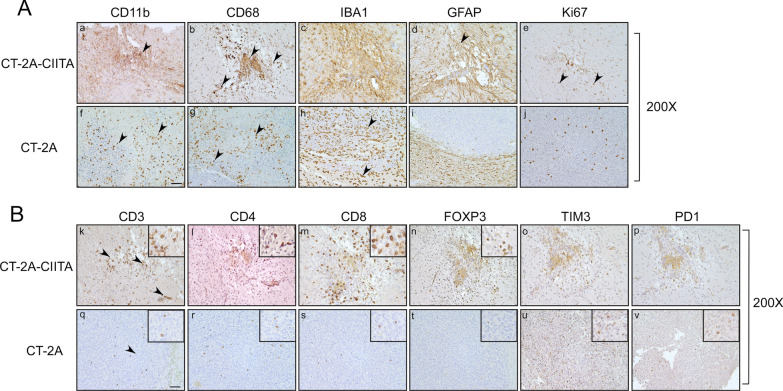
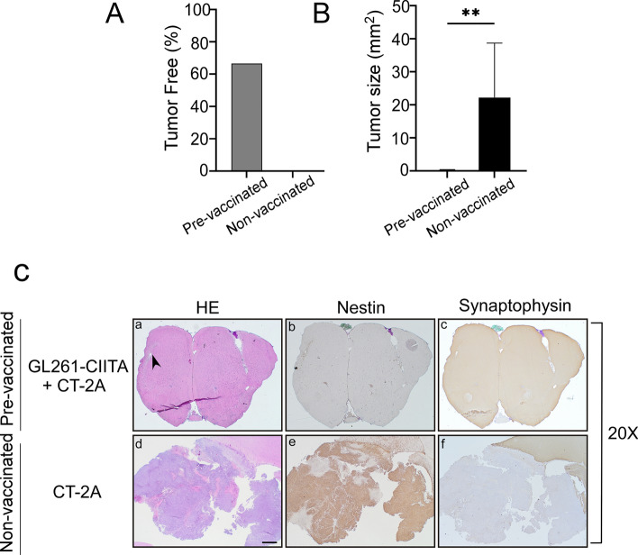
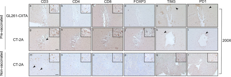
Methods: To assess tumor growth and immune response, immunocompetent mice were injected intracranially with either CT-2A or CIITA-modified CT-2A (CT-2A-CIITA) cells, and brain tissues were examined histologically. Additionally, mice vaccinated with GL261-CIITA cells were subsequently challenged with parental CT-2A cells in the opposite hemisphere, followed by immunohistochemical analysis of brain tissues.

Results: Results indicated that CT-2A-CIITA cells were either rejected or exhibited delayed tumor growth in immunocompetent mice. Furthermore, mice previously vaccinated with GL261-CIITA cells demonstrated efficient rejection of CT-2A tumors, suggesting the induction of cross-protective immunity. This immune response was associated with a shift from an immunosuppressive to an immunogenic tumor microenvironment, implying the development of adaptive immunity against shared tumor antigens.

Conclusions: In conclusion, these findings support the broader applicability of CIITA-based vaccination against GBM and provide the first evidence of shared immunogenic antigens between two distinct murine GBM models. This discovery opens avenues for identifying common tumor antigens through immunopeptidomics, which could inform future immunotherapeutic strategies for glioblastoma.

Keywords: CT-2A; Cross-protection; Glioblastoma; Immunotherapy; Tumor associated antigens; Vaccine.


Conflict of interest statement

Declarations. Competing interests: All authors declare that no conflicts of interest exist.

Figures

Fig. 1
Fig. 1
Intracranial implantation of MHC-II positive CT-2A-CIITA tumor cells results in tumor rejection in vivo. C57BL/6 mice received intracranial injection of 1 × 104 CT-2A or CT-2A-CIITA glioma cells. On day 21 after injection, mice were sacrificed, brains were removed, and serial sections of the brain were carried out to measure tumor size and for IHC staining. A Percentage of tumor free mice, N = 18/group. B HE and IHC staining of serial brain sections. Representative histological sections of the brains harvested from mice injected with CT-2A-CIITA and rejecting the tumor or with CT-2A. The first two series of upper panels were taken at ×20 magnification; scale bar corresponds to 500 μM. The third and fourth series of horizontal panels were taken at ×200 magnification; scale bar corresponds to 50 μM. Small square boxes are the areas represented in the corresponding large square boxes of each IHC image. Small square boxes were taken at ×400 magnification. Slides from the brain tissues isolated from CT-2A or CT-2A-CIITA tumor bearing mice were subjected to immunohistochemical staining with anti-Nestin, anti-Synaptophysin and anti-MHC-II antibodies. HE haematoxylin and eosin, IHC immunohistochemical staining
Fig. 2
Fig. 2
Immunohistological characterization of CT-2A-CIITA tumors compared to CT-2A control group. A Representative histological brain sections harvested from mice injected with CT-2A-CIITA (upper panels) or with CT-2A (bottom panels) at ×200 magnification. Scale bar corresponds to 50 μM. Slides from the brain tissues isolated from CT-2A or CT-2A-CIITA tumor bearing mice were subjected to immunostaining with anti-CD11b, anti-CD68, anti-IBA1, anti-GFAP, and anti-Ki67 antibodies. Arrowhead points to cells with corresponding relevant marker whose characteristics are described in the Result section. B Immunostaining for CD3, CD4, CD8, FOXP3, PD1 and TIM3 in CT-2A (upper panels) and CT-2A-CIITA (bottom panels) at ×200 magnification. Scale bar corresponds to 50 μM. Small square boxes are the areas represented in the corresponding large square boxes of each IHC image. Small square boxes were taken at ×400 magnification. Note that selected areas in IHC images of CT-2A parental tumors were taken in the rare zones in which positive cells for the selective marker were present. IHC, immunohistochemistry. Arrowheads point to cells with the relevant marker whose characteristics are described in “Results” section
Fig. 3
Fig. 3
Preventive vaccination with GL261-CIITA tumor cells protects the animal against challenge with CT-2A parental tumor cells. C57BL/6 mice were intracranially (i.c.) injected with GL261-CIITA cells into the right striatum and after 21 days challenged with parental CT-2A tumor cells in the left striatum (pre-vaccinated), N = 9. After 3 additional weeks, animals were sacrificed, and their brains were analyzed histologically for presence and size of tumors. As a control, another group of mice were i.c. injected with CT-2A cells (non-vaccinated), and their brains were analyzed after 3 weeks, N = 9. A Percentage of tumor free mice, N = 9. B Average size of CT-2A tumors in pre-vaccinated and non-vaccinated mice. Bars represent mean values, and error bars indicate the SD of each group. p-Values were determined via unpaired t-test; **p < 0.01. C Representative histological brain sections harvested from pre-vaccinated mice injected with GL261-CIITA (panels a–c) and with CT-2A (panels d–f), at ×20 magnification. Scale bar corresponds to 50 μM. Sections were stained with HE or by IHC with Nestin- and Synaptophysin-specific antibodies to better identify tumoral and non-tumoral tissue, respectively. HE hematoxylin and eosin
Fig. 4
Fig. 4
Immunohistological characterization of CT-2A tumor rejection in GL261-CIITA pre-vaccinated mice. Representative histological brain sections harvested from pre-vaccinated mice (N = 9) injected with GL261-CIITA (panels a–e) and with CT-2A (panels f–j), and from non-vaccinated mice injected with CT-2A only (N = 9) (panels: k–o) at ×200 magnification. Scale bar corresponds to 500 μM. Slides from the brain tissues isolated from pre-vaccinated and non-vaccinated tumor bearing mice were subjected to immunohistochemical staining with anti-Nestin, anti-Synaptophysin, anti-MHC-II and anti-Ki67 antibodies
Fig. 5
Fig. 5
Immunohistological characterization of CT-2A tumor microenvironment in pre-vaccinated mice. Representative histological brain sections harvested from pre-vaccinated mice (N = 9) injected with GL261-CIITA (panels a–d) and with CT-2A (panels e–h), and from non-vaccinated mice injected with CT-2A only (N = 9) (panels i–l) at ×200 magnification. Scale bar corresponds to 500 μM. Small square boxes are the areas represented in the corresponding large square boxes of each IHC image. Small square boxes were taken at ×400 magnification. Slides from the brain tissues isolated from pre-vaccinated and non-vaccinated tumor bearing mice were subjected to immunohistochemical staining with anti-CD11b, anti-CD68, anti-IBA1 and anti-GFAP
Fig. 6
Fig. 6
Rejected CT-2A parental tumors in pre-vaccinated mice are strongly infiltrated by anti-tumor T cell cells. Slides from the brain tissues were subjected to immunohistochemical staining with anti-CD3, anti-CD4, anti-CD8, anti-FOXP3, anti-PD1 and anti- TIM3 antibodies. Small square boxes are the areas represented in the corresponding large square boxes of each IHC image. Images were taken at ×200 magnification. Scale bar corresponds to 500 μM. Large square boxes were taken at ×400 magnification. Note that selected areas in IHC images of CT-2A parental tumors of non-vaccinated mice are taken in the rare zones in which positive cells for the selective marker were present. The lymphocytic compartment was primarily distributed throughout challenged CT-2A tumor bed (black arrowhead). In the control non-vaccinated mice, only a scattered and a minimal presence of T-lymphocytes was observed within the tumor mass (arrowheads). Some TIM3 + and PD1 + tumor cells were observed in CT-2A tumors (small square boxes)

Similar articles

References

    1. Stupp R, Mason WP, van den Bent MJ, Weller M, Fisher B, Taphoorn MJB, et al. Radiotherapy plus concomitant and adjuvant temozolomide for glioblastoma. N Engl J Med. 2005;352:987–96. - PubMed
    1. Miranda-Filho A, Piñeros M, Soerjomataram I, Deltour I, Bray F. Cancers of the brain and CNS: global patterns and trends in incidence. Neuro-Oncol. 2017;19:270–80. - PMC - PubMed
    1. Preusser M, de Ribaupierre S, Wöhrer A, Erridge SC, Hegi M, Weller M, et al. Current concepts and management of glioblastoma. Ann Neurol. 2011;70:9–21. - PubMed
    1. Qi D, Li J, Quarles CC, Fonkem E, Wu E. Assessment and prediction of glioblastoma therapy response: challenges and opportunities. Brain J Neurol. 2023;146:1281–98. - PMC - PubMed
    1. Huang AC, Zappasodi R. A decade of checkpoint blockade immunotherapy in melanoma: understanding the molecular basis for immune sensitivity and resistance. Nat Immunol. 2022;23:660–70. - PMC - PubMed