Review
. 2025 Sep;247(6):2538-2556.
doi: 10.1111/nph.70370. Epub 2025 Jul 8.

Evolution of petal patterning: blooming floral diversity at the microscale

Affiliations
Review

Evolution of petal patterning: blooming floral diversity at the microscale

Erin Doody et al. New Phytol. 2025 Sep.

Abstract

The flowers of angiosperms are extraordinarily diverse. While most floral variation is visible to the naked eye, this diversity goes beyond the macroscale: Floral organs comprise an underappreciated range of cell types that generate a multitude of patterns across their surfaces and give rise to novel structures. Because diverse cell patterns provide adaptations to biotic and abiotic factors, they also contribute to angiosperm evolution and speciation. Yet, how such diversity originates remains to be understood. In this review, we focus on petals, which together form the corolla, to examine the mechanisms patterning floral surfaces at the cellular level. We summarize current research aiming to understand how cell fate specification and controlled cell growth (proliferation and expansion) are achieved with high spatial resolution during petal development. We also examine the adaptive potential for such patterns and how they contribute to plant fitness and diversification. Finally, we discuss promising directions for future research on the evolution of petal patterning at the microscale and identify outstanding questions that technological advances now make it possible to address.

Keywords: cell type; corolla; evo‐devo; floral development; natural variation; novelty; petal patterning; pollinator attraction.


Conflict of interest statement

None declared.

Figures

Fig. 1
Fig. 1
Model systems currently used to study flower evo‐devo at the microscale. (a) Brachypodium distachyon, (b) Phalaenopsis moth orchid, (c) Nigella sp., (d) Eschscholzia californica, (e) Aquilegia coerulea sp., (f) Delphinium sp., (g) Hibiscus trionum, (h) Clarkia amoena, (i) Mimulus guttatus, (j) Antirrhinum majus, (k) Petunia × hybrida and (l) Daucus carota. (m) Simplified phylogenetic tree of the angiosperms according to the APGIV classification (The Angiosperm Phylogeny Group et al., 2016), depicting the distribution of these model species. Pictures in (a, l) are credited to Laval University and Harry Rose, respectively, via Wikimedia Commons.
Fig. 2
Fig. 2
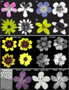
Differences in ultraviolet (UV) light reflecting and absorbing pigments as well as cell shape and texture across the petal surface can generate petal patterns in the UV range. (a–d) Images of flowers in human‐visible and UV light spectrums of (a) Pelargonium ‘Pink capitatum (Geraniaceae), (b) Geranium sylvaticum (Wood Cranes‐Bill, Geraniaceae), (c) Rudbeckia laciniata (Cut‐leaf coneflower, Asteraceae) and (d) Bidens triplinervia (Asteraceae), each with variation in UV‐absorbing or reflective properties across the corolla. (e) Natural variation in petal pigmentation patterns and UV‐reflective properties of Coreopsis tunctoria (Nuttall Weed, Asteraceae), with variation in UV reflection varying with both pigment and cell texture. (f) Artificial epoxy resin flowers created by imprinting the petal of Hibiscus trionum in dental wax to replicate its surface: striated flat elongated cells in the proximal portion and smooth conical cells in the distal portion of the petal, as seen under SEM imaging (Top left: SEM image of distal smooth conical cells; Bottom left; SEM image of proximal flat striated cells). In the purple artificial flower (left), the conical cells create a mat velvety appearance on the corolla periphery while the flat striated cells render its centre shiny. This ‘structural’ bullseye is not easily visible to the human eye in the white artificial flower (middle) but become apparent when imaged with a UV camera (right), showing that pigment, shape and texture of epidermal cells all influence visible and UV light reflective properties of the petal.
Fig. 3
Fig. 3
Micropatterns on the petal surface are created by variation in epidermal cell pigment, shape and cuticle textures. (a) Patterns on the petal of Erodium castellanum (Geraniaceae) created by cell‐specific pigmentation. (b) Variation in cell shape across the petals of Hardenbergia violaceae (Fabaceae). (c) Smooth cuticle on elongated petal epidermal cells of Ceratostigma griffithii (Plumbaginaceae). (d) Wrinkled cuticle on the elongated petal epidermal cells of Gaura lindheimeri (Onagraceae). (e) Smooth cuticle on the conical petal epidermal cells of Hibiscus trionum (Malvaceae). (f) Wrinkled cuticle on the conical petal epidermal cells of Dahlia merckii (Asteraceae).
Fig. 4
Fig. 4
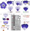
MYB transcription factors are major contributors to petal pattern development and evolution. (a) Spatial restriction of MYB gene expression (e.g. subgroup 6 (SG6) R2R3‐myeloblastosis (MYB) promote anthocyanin production while subgroup 7 (SG7) R2R3‐MYB activate flavonol synthesis) largely accounts for pigmentation pattern formation during petal development; (b) variation in expression domain or (c) in the identity of the MYB gene expressed in a given region (subgroup 21 R2R3‐MYB can promote the production of yellow carotenoids) can generate the intra‐ and interspecific diversity in petal patterns. (d) Variation in pattern proportions can be due to mutation affecting the coding sequence or the regulatory region of MYB genes. Red crosses depict deleterious mutations, and green crosses represent gain of cis‐regulatory elements. (e) Petal pigmentation spots can emerge from the interactions between a transcriptional activator (SG6 R2R3‐MYB) and a repressor (e.g. R3‐MYB). Green arrows indicate transcriptional activation, red blunt arrow indicate transcriptional repression. (f) Post‐transcriptional processes targeting SG6 R2R3‐MYBs can also yield petal pattern variation, reducing pigment production via changes in the 5′UTR impacting protein translation (right) or abolishing pigment production in certain petal domains via siRNA promoting the degradation of SG6 R2R3‐MYB transcripts (left). Arrows indicate transcription and translation steps. (g) MYB genes are also involved in specifying cell shape and cuticular texture, but many outstanding questions remain: Beyond bHLH/WD40 that help MYB regulate anthocyanin production and AP2/ERF factors that participate in cuticular ridges formation, most of the molecular players working along MYBs to control epidermis cell characteristics are yet to be identified. What specifies the geometry of nonconical cell shape and what decides whether SG9 MYBs induce conical cell or trichome formation is also not understood. (h) Crucially, the signalling events and upstream regulators that specify petal polarity, pattern cell fate along the axes of developing petal primordia and divide the petal surface into distinct territories where growth can be controlled independently, and neighbouring cells can acquire contrasting fates remain unclear. Brown arrows indicate the developmental progression from emerging primordia on the floral meristem to mature petals in open flowers.
Fig. 5
Fig. 5
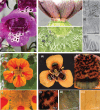
Variation in trichome‐like structures creates floral micropatterns. (a) Digitalis purpurea (Foxglove) with elongated trichomes on its corolla (i). (b) Three trichome domains in the floral nectary of Hibiscus trionum. (i) Nectar‐secreting trichomes closest to the base of the flower, (ii) single‐celled defensive trichomes in the middle region and (iii) enlarged glandular trichomes at the periphery of the nectary. (c) The petal of Tropaeolum majus (Nasturtium) with (i) serration‐like outgrowths. (d) The petal of Moraea tulbaghensis with different epidermal cell types in three discrete domains (i). (ii) Cilia‐like structures in the proximal region, (iii) iridescent conical cells in the middle region and (iv) elongated yellow cells at the distal region of the petal.
Fig. 6
Fig. 6
Individuation and change in growth at the cellular scale can support the evolution of novelty and biodiversity. (a) Nectar spur specification allows a subdomain of the petal primordia to become differentiated (individuation) and a spur emerges as cell proliferation is promoted in this ‘spur domain’. Depending on the genus, spur elongation is mostly driven through cell proliferation or cell expansion. (b) Interspecific variation in spur length is due to changes in cell expansion (differences in cell length/anisotropy) in Aquilegia (Puzey et al., 2012) and variation in cell proliferation (no difference in cell length but difference in number of cells) in Linaria (Cullen et al., 2018). Red arrows depict the average cell length in each case (c) Spur formation (black arrow) can be induced in spurless species, such as snapdragon (another species of Lamiales) or tobacco (Solanales), when a KNOX transcription factor promoting cell growth is constitutively overexpressed (Box et al., 2011).

Similar articles

References

    1. Abrahamczyk S, Humphreys AM, Trabert F, Droppelmann F, Gleichmann M, Krieger V, Linnartz M, Lozada‐Gobilard S, Rahelivololona ME, Schubert M et al. 2021. Evolution of brood‐site mimicry in Madagascan impatiens (Balsaminaceae). Perspectives in Plant Ecology, Evolution and Systematics 49: 125590.
    1. Airoldi CA, Lugo CA, Wightman R, Glover BJ, Robinson S. 2021. Mechanical buckling can pattern the light‐diffracting cuticle of Hibiscus trionum . Cell Reports 36: 109715. - PMC - PubMed
    1. Alonso‐Blanco C, Andrade J, Becker C, Bemm F, Bergelson J, Borgwardt KM, Cao J, Chae E, Dezwaan TM, Ding W et al. 2016. 1,135 genomes reveal the global pattern of polymorphism in Arabidopsis thaliana . Cell 166: 481–491. - PMC - PubMed
    1. Amrad A, Moser M, Mandel T, de Vries M, Schuurink RC, Freitas L, Kuhlemeier C. 2016. Gain and loss of floral scent production through changes in structural genes during pollinator‐mediated speciation. Current Biology 26: 3303–3312. - PubMed
    1. Antoniou Kourounioti RL, Band LR, Fozard JA, Hampstead A, Lovrics A, Moyroud E, Vignolini S, King JR, Jensen OE, Glover BJ. 2013. Buckling as an origin of ordered cuticular patterns in flower petals. Journal of the Royal Society Interface 10: 20120847. - PMC - PubMed

LinkOut - more resources