

Combining radiomics and deep learning to predict liver metastasis of gastric cancer on CT image
- PMID: 40630210
- PMCID: PMC12234316
- DOI: 10.3389/fonc.2025.1613972
Combining radiomics and deep learning to predict liver metastasis of gastric cancer on CT image
Abstract
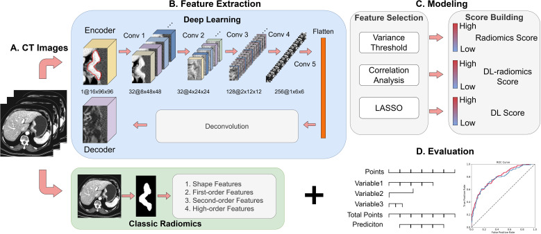
Objective: Our study aimed to explore the potential of deep learning (DL) radiomics features from CT images of primary gastric cancer (GC) in predicting gastric cancer liver metastasis (GCLM) by establishing and verifying a prediction model based on clinical factors, classical radiomics and DL features.
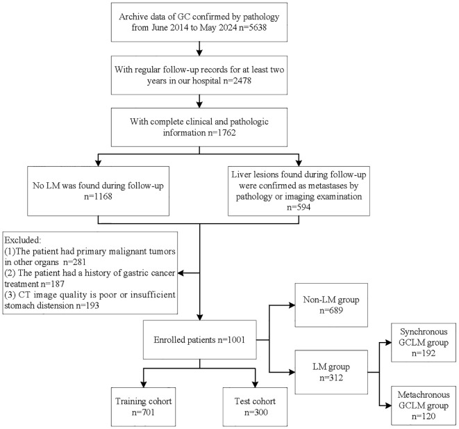
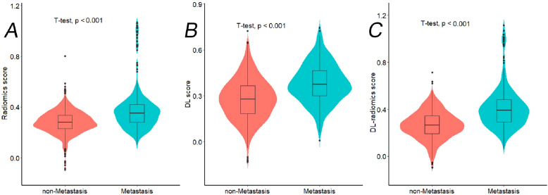
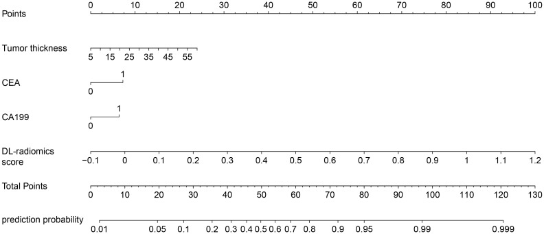
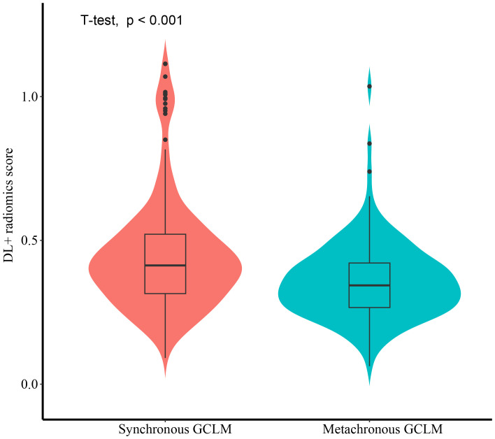
Methods: We retrospectively analyzed 1001 pathologically confirmed GC patients from June 2014 to May 2024, divided into non-LM (n=689) and LM groups (n=312). CT-based classic radiomics and DL features were extracted and screened to construct a DL-radiomics score. This score, along with statistically significant clinical factors, was used to build a fused model which visualized as a nomogram. The model's predictive performance, calibration, and clinical utility were assessed and compared against a clinical model. Additionally, the DL-radiomics score's role in distinguishing between synchronous and metachronous GCLM was evaluated.
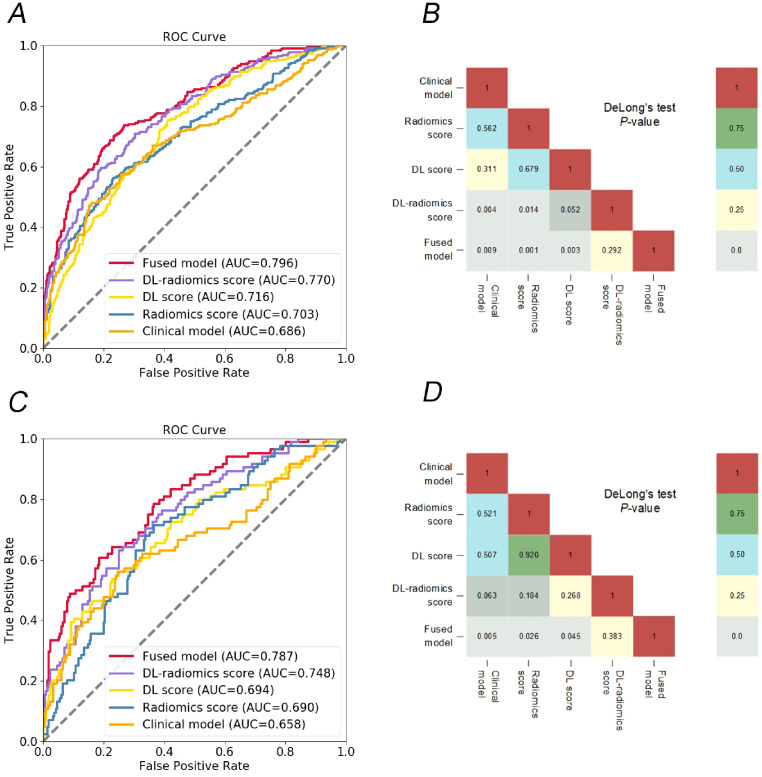
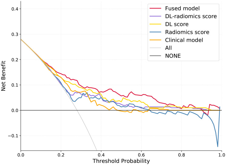
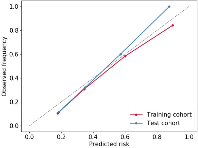
Results: The fused model showed good predictive performance [AUC: 0.796 (95% CI: 0.766-0.826) in training cohort and 0.787 (95% CI: 0.741-0.834) in test cohort], outperforming the clinical model, radiomics score and DL score (P<0.05). In addition, the decision curve confirmed that the model provided the largest clinical net benefit compared with all other models in the relevant threshold. DL-radiomics score showed moderate predictive performance in distinguishing between synchronous GCLM and metachronous GCLM, with an AUC of 0.665 (95% CI, 0.613-0.718).
Conclusion: The CT-based fused model has demonstrated significant value in predicting the occurrence of GCLM, and can provide a reference for the personalized follow-up and treatment of patients.
Keywords: computed tomography; deep learning; gastric cancer; liver metastasis; radiomics nomogram.
Copyright © 2025 Guo, Yin, Zhang, Liang, Gao and Cheng.
Conflict of interest statement
The authors declare that the research was conducted in the absence of any commercial or financial relationships that could be construed as a potential conflict of interest.
Figures
Similar articles
-
Are Current Survival Prediction Tools Useful When Treating Subsequent Skeletal-related Events From Bone Metastases?Clin Orthop Relat Res. 2024 Sep 1;482(9):1710-1721. doi: 10.1097/CORR.0000000000003030. Epub 2024 Mar 22. Clin Orthop Relat Res. 2024. PMID: 38517402
-
Deep learning radiomics nomogram for preoperatively identifying moderate-to-severe chronic cholangitis in children with pancreaticobiliary maljunction: a multicenter study.BMC Med Imaging. 2025 Feb 5;25(1):40. doi: 10.1186/s12880-025-01579-3. BMC Med Imaging. 2025. PMID: 39910477 Free PMC article.
-
Preoperative prediction value of 2.5D deep learning model based on contrast-enhanced CT for lymphovascular invasion of gastric cancer.Sci Rep. 2025 Jul 15;15(1):25646. doi: 10.1038/s41598-025-11427-7. Sci Rep. 2025. PMID: 40664824 Free PMC article.
-
MRI-Based Radiomics Methods for Predicting Ki-67 Expression in Breast Cancer: A Systematic Review and Meta-analysis.Acad Radiol. 2024 Mar;31(3):763-787. doi: 10.1016/j.acra.2023.10.010. Epub 2023 Nov 2. Acad Radiol. 2024. PMID: 37925343
-
Cost-effectiveness of using prognostic information to select women with breast cancer for adjuvant systemic therapy.Health Technol Assess. 2006 Sep;10(34):iii-iv, ix-xi, 1-204. doi: 10.3310/hta10340. Health Technol Assess. 2006. PMID: 16959170
References
LinkOut - more resources
Full Text Sources
Research Materials
Miscellaneous