Case Report: A heterozygous loss-of-function variant of the ERG gene in a family with vascular pathologies
- PMID: 40630904
- PMCID: PMC12235746
- DOI: 10.3389/fcvm.2025.1550523
Case Report: A heterozygous loss-of-function variant of the ERG gene in a family with vascular pathologies
Abstract
Background: The transcription factor ERG (erythroblast transformation-specific-related gene) has been identified as a key regulator of vascular function by suppressing inflammation in endothelial cells (ECs). Dysregulation of ERG due to genetic risk variants is linked to chronic inflammation in conditions such as atherosclerosis and aortic aneurysms.
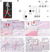
Case presentation: This research work investigates the role of the ERG gene in the development of a systemic arterial aneurysm manifestation. Given the previous implication of ERG in vascular development, we now report a loss-of-function variant (Leu212*) in the ERG gene, segregating in a family with vascular pathologies. Multiple arterial aneurysms were observed in one family member, and early onset of vascular-associated stroke in another individual carrying the familial ERG variant. Histological analysis of arterial aneurysm specimen showed comparable expression of ERG in endothelial cells of the vasa vasorum in samples from the patient and controls.
Conclusion: Our report discusses the possibility that loss-of-function variants in ERG may act as a risk factor for arterial disease.
Keywords: ERG; abdominal aortic aneurysm; haploinsufficiency; loss-of-function variant; multiple arterial aneurysms.
© 2025 Erhart, Dikow, Schwaibold, Dihlmann, Grond-Ginsbach, Körfer, Schaaf, Oeser, Hinderhofer, Böckler, Zerella, Scott, Hahn and Marbach.
Conflict of interest statement
The authors declare that the research was conducted in the absence of any commercial or financial relationships that could be construed as a potential conflict of interest.
Figures


Similar articles
-
Drugs to reduce bleeding and transfusion in major open vascular or endovascular surgery: a systematic review and network meta-analysis.Cochrane Database Syst Rev. 2023 Feb 17;2(2):CD013649. doi: 10.1002/14651858.CD013649.pub2. Cochrane Database Syst Rev. 2023. PMID: 36800489 Free PMC article.
-
Endovascular treatment for ruptured abdominal aortic aneurysm.Cochrane Database Syst Rev. 2017 May 26;5(5):CD005261. doi: 10.1002/14651858.CD005261.pub4. Cochrane Database Syst Rev. 2017. PMID: 28548204 Free PMC article.
-
Totally percutaneous versus surgical cut-down femoral artery access for elective bifurcated abdominal endovascular aneurysm repair.Cochrane Database Syst Rev. 2023 Jan 11;1(1):CD010185. doi: 10.1002/14651858.CD010185.pub4. Cochrane Database Syst Rev. 2023. PMID: 36629152 Free PMC article.
-
Signs and symptoms to determine if a patient presenting in primary care or hospital outpatient settings has COVID-19.Cochrane Database Syst Rev. 2022 May 20;5(5):CD013665. doi: 10.1002/14651858.CD013665.pub3. Cochrane Database Syst Rev. 2022. PMID: 35593186 Free PMC article.
-
Interventions for central serous chorioretinopathy: a network meta-analysis.Cochrane Database Syst Rev. 2025 Jun 16;6(6):CD011841. doi: 10.1002/14651858.CD011841.pub3. Cochrane Database Syst Rev. 2025. PMID: 40522203
References
-
- Li Y, Ren P, Dawson A, Vasquez HG, Ageedi W, Zhang C, et al. Single-cell transcriptome analysis reveals dynamic cell populations and differential gene expression patterns in control and aneurysmal human aortic tissue. Circulation . (2020) 142(14):1374–88. 10.1161/CIRCULATIONAHA.120.046528 - DOI - PMC - PubMed
Publication types
LinkOut - more resources
Full Text Sources

