. 2025 Jul 1:2025:3678453.
doi: 10.1155/joph/3678453. eCollection 2025.

Individual Risk Assessment and Prognostication of Outcomes After Corneal Cross-Linking

Affiliations

Individual Risk Assessment and Prognostication of Outcomes After Corneal Cross-Linking

Y Statsenko et al. J Ophthalmol. .

Abstract

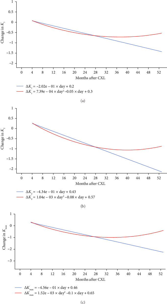
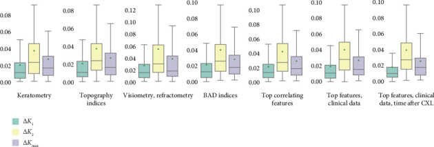
Background and Objective: Corneal collagen cross-linking (CXL) is a treatment which arrests keratoconus (KC) progression, but its effectiveness differs radically among patients. Herein, we report preoperative diagnostic findings that reflect CXL outcomes and allow physicians to prognosticate treatment efficiency. Methods: In a medical centre, we retrospectively analysed pre- and postoperative data about 107 patients (112 eyes) treated with CXL from January 2018 to December 2022. Exclusion criteria were age below 16 years, a corneal thickness below 400 microns, severe dry eye, other corneal diseases/infections, re-CXL, pregnancy and missing follow-up examinations. All the subjects (79 males and 28 females) were followed for a minimum of 4 and a maximum of 40 months. The study dataset was comprised of 796 cases of clinical assessment, pachymetry, visiometry, refractometry and topography examinations. With these data, we modelled maximum anterior keratometry (K max) and curvature power of the flat and steep meridians of the corneal anterior surface (K 1 and K 2). Results: Two years after the invasion, corneal curvature coefficients decreased progressively. Then, they remained stable for four months and rose afterwards. In the most accurate K 1, K 2 and K max models, the proportion of mean absolute error to the range of values was 1.72, 3.66 and 2.37%, respectively. Pronounced corneal thinning, low best-corrected visual acuity and high K max levels predict unfavourable outcomes. Conclusions: The high accuracy of the models advocates for a personalised approach to candidate selection for CXL.

Keywords: corneal collagen cross-linking; explainable artificial intelligence; intervention outcome; keratoconus; keratometry readings; machine learning; precision medicine; predictive model.


Conflict of interest statement

The authors declare no conflicts of interests.

Figures

Figure 1
Figure 1
Schematic presentation of exclusion map and zone on corneal surface.
Figure 2
Figure 2
Coefficients of Pearson correlations between postoperative changes in K1, K2, Kmax and preoperative values of these parameters (p < 0.05).
Figure 3
Figure 3
Coefficients of Pearson correlations between flat, steep and maximum anterior keratometry before and after CXL (p < 0.05).
Figure 4
Figure 4
Pearson correlations of postoperative keratometry readings with preoperative keratometry, pachymetry, visiometry, refractometry findings (a), corneal topography and deviation indices (b). Text colour encodes p level; weak associations (p > 0.05) are crossed out.
Figure 5
Figure 5
Pearson correlations of postoperative flat, steep and maximum keratometries with preoperative parameters of elevation back maps: corneal thickness (a), posterior elevation (b), posterior elevation with best-fit sphere (c), posterior elevation excluding thinnest zone (d) and excluded thinnest zone of posterior elevation (e). Numbers denote r-values of strong correlations (p < 0.05); heatmap colour indicates p level.
Figure 6
Figure 6
Postoperative changes in flat (a), steep (b) and maximum keratometries (c).
Figure 7
Figure 7
Performance of regression models predicting change in K1, K2 and Kmax.

Similar articles

References

    1. Gomes J. A. P., Rodrigues P. F., Lamazales L. L. Keratoconus Epidemiology: A Review. Saudi Journal of Ophthalmology . 2022;36(1):3–6. doi: 10.4103/sjopt.sjopt_204_21. - DOI - PMC - PubMed
    1. Kristianslund O., Drolsum L. Prevalence of Keratoconus in Persons with Down Syndrome: A Review. BMJ Open Ophthalmology . 2021;6(1):p. e000754. doi: 10.1136/bmjophth-2021-000754. - DOI - PMC - PubMed
    1. Syed Z. A., Tomaiuolo M., Zhang Q., et al. Trends and Sociodemographic Patterns in Keratoconus Management 2015–2020: An American Academy of Ophthalmology Iris® Registry Analysis. Ophthalmology . 2024;131(8):892–901. doi: 10.1016/j.ophtha.2024.01.036. - DOI - PubMed
    1. Santodomingo-Rubido J., Carracedo G., Suzaki A., Villa-Collar C., Vincent S. J., Wolffsohn J. S. Keratoconus: An Updated Review. Contact Lens and Anterior Eye . 2022;45(3):p. 101559. doi: 10.1016/j.clae.2021.101559. - DOI - PubMed
    1. Hashemi H., Heydarian S., Hooshmand E., et al. The Prevalence and Risk Factors for Keratoconus: a Systematic Review and Meta-Analysis. Cornea . 2020;39(2):263–270. doi: 10.1097/ico.0000000000002150. - DOI - PubMed

LinkOut - more resources