. 2025 Jun 24:13:1576809.
doi: 10.3389/fbioe.2025.1576809. eCollection 2025.

Investigation of corneal hydration and the impact of cross-linking therapy on water retention using Brillouin spectroscopy, Raman spectroscopy and polarization-sensitive optical coherence tomography

Affiliations

Investigation of corneal hydration and the impact of cross-linking therapy on water retention using Brillouin spectroscopy, Raman spectroscopy and polarization-sensitive optical coherence tomography

Jan Rix et al. Front Bioeng Biotechnol. .

Abstract

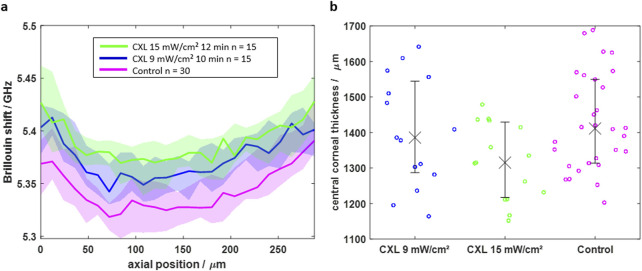
Recently, Brillouin spectroscopy has been proposed as a promising non-invasive tool to evaluate corneal biomechanics, e.g., during corneal cross-linking (CXL) treatment. However, the impact of corneal hydration on the Brillouin shift hampers straightforward interpretation of the measurements, especially when judging on the success of the CXL procedure. Therefore, in this work, we first quantify the effect of corneal (de)hydration on the Brillouin shift revealing that reliable measurements are only possible under constant hydration conditions, which was subsequently achieved by immersing porcine eyes in solution and waiting until an equilibrium state was reached. Investigations showed that Brillouin spectroscopy evaluates the CXL effect mainly indirectly via reduced water uptake, while polarization-sensitive optical coherence tomography evaluates the CXL effect directly via changes in collagen fiber alignment and is therefore insensitive to corneal hydration. Raman spectroscopy is not indicating any alterations in the molecular structure revealing that new cross-links are not created due to the CXL procedure. Compared to large water retention in balanced salt solution, the missing water uptake in dextran-based (16%) solution hampers the evaluation of the CXL effect by Brillouin spectroscopy.

Keywords: Brillouin; PS-OCT; Raman; cornea; corneal cross-linking; hydration.


Conflict of interest statement

RH and RK received lecture fees from Oculus Optikgeraete GmbH (Wetzlar, Germany) and Heidelberg Engineering GmbH (Heidelberg, Germany). The remaining authors declare that the research was conducted in the absence of any commercial or financial relationships that could be construed as a potential conflict of interest.

Figures

FIGURE 1
FIGURE 1
(a) Brillouin/Raman microscope setup with eyes being measured in solution via a dipping objective. While laser light (red) is filtered out by Rubidium cells, Brillouin scattered light (purple dashed) of the sample and the methanol reference signal is analyzed by a 2-stage VIPA spectrometer and Raman scattered light (orange dashed) is analyzed with a commercial grating-based spectrometer. (b) Picture of the setup, where the eye is totally immersed in solution.
FIGURE 2
FIGURE 2
(a) PS-OCT setup consisting of the base unit, the beam scanning system and the scan lens kit. (b) Picture of the eye being placed in solution and measured through a cover slip added to the objective.
FIGURE 3
FIGURE 3
Time-dependence of dehydration process after de-epithelization showing that (a) the Brillouin shift increases and (b) the central corneal thickness decreases due to evaporation. Dashed colored curves are for individual eyes, whereas black solid curve is the median curve and the grey area the interquartile range, respectively.
FIGURE 4
FIGURE 4
Time-dependent measurements after immersing eyes in BSS showing that (a) the Brillouin shift decreases and (b) the CCT increases over time. The Brillouin shift reaches an equilibrium, whereas the CCT still increases after 27 min. Dashed colored curves are for individual eyes, whereas black solid curve is the median curve and the grey area the interquartile range, respectively.
FIGURE 5
FIGURE 5
(a) Axial BS scans of CXL-treated (blue and green) and control (magenta) eyes indicating that CXL results in a higher Brillouin shift in the first 264 μm. (b) The central corneal thickness shows lower values for CXL-treated eyes, which is attributed to a mitigated water uptake. Solid line/crosses and shaded area/error bars are indicating the median value and interquartile range, respectively.
FIGURE 6
FIGURE 6
Mean Raman spectra of CXL-treated (blue and green) and control eyes (magenta), indicating that CXL does not lead to significant changes in Raman spectra. In particular, the strong signals (dashed lines) show no noticeable changes, which is an indicator of an unchanged molecular structure.
FIGURE 7
FIGURE 7
(a) DOP over the normalized depth showing that CXL-treated eyes have an increased DOP in the region of 0–0.15 relative depth. (b) Scatter plot of the mean DOP values in this region for all individual eyes indicating the statistical significance of the CXL effect on the DOP (*p < 0.05, **p < 0.01).
FIGURE 8
FIGURE 8
(a) DOP over the normalized depth showing that CXL-treated eyes (cyan) in 16% Dextran solution have an increased DOP in the region of 0–0.28 relative depth compared to control eyes (red). (b) Scatter plot of the mean DOP values in this region for individual eyes indicating the statistical significance of the CXL effect on the DOP (*p < 0.05).

References

    1. Abass A., Hayes S., White N., Sorensen T., Meek K. M. (2015). Transverse depth-dependent changes in corneal collagen lamellar orientation and distribution. J. R. Soc. Interface 12 (104), 20140717. 10.1098/rsif.2014.0717 - DOI - PMC - PubMed
    1. Al-Mujaini A., Wali U. K., Azeem S. (2013). Optical coherence tomography: clinical applications in medical practice. Oman Med. J. 28 (2), 86–91. 10.5001/omj.2013.24 - DOI - PMC - PubMed
    1. Antonacci G., Beck T., Bilenca A., Czarske J., Elsayad K., Guck J., et al. (2020). Recent progress and current opinions in Brillouin microscopy for life science applications. Biophys. Rev. 12 (3), 615–624. 10.1007/s12551-020-00701-9 - DOI - PMC - PubMed
    1. Bao Q., Newman B., Wang Y., Choi S., Burgess D. J. (2018). In vitro and ex vivo correlation of drug release from ophthalmic ointments. J. Control. Release 276 (April), 93–101. 10.1016/j.jconrel.2018.03.003 - DOI - PMC - PubMed
    1. Baumann B., Augustin M., Lichtenegger A., Harper D. J., Muck M., Eugui P., et al. (2018). Polarization-sensitive optical coherence tomography imaging of the anterior mouse eye. J. Biomed. Opt. 23 (8), 1. 10.1117/1.JBO.23.8.086005 - DOI - PubMed

LinkOut - more resources