. 2025 Jun 24:16:1618650.
doi: 10.3389/fimmu.2025.1618650. eCollection 2025.

Applications and insights from continuous dengue virus infection in a stable cell line

Affiliations

Applications and insights from continuous dengue virus infection in a stable cell line

M Jane Morwitzer et al. Front Immunol. .

Abstract

Dengue is caused by the four serotypes of dengue virus (DENV-1-4) and poses a significant global public health challenge, with an estimated 100-400 million infections annually. Severe dengue manifestations, such as Dengue Hemorrhagic Fever (DHF) and Dengue Shock Syndrome (DSS), are influenced by immune responses, particularly during secondary infections with different serotypes. Antibody-dependent enhancement (ADE) of DENV infection is a critical mechanism in dengue immunopathogenesis, underscoring the need for comprehensive evaluation of antibody responses. Traditional cell lines used for DENV propagation exhibit variability and present logistical challenges for assessing non-neutralizing antibody functions. Here, we report the establishment of a stable CEM-NKR cell line expressing DC-SIGN, designated CEM2001, capable of supporting continuous infection with all four DENV serotypes. These cell lines allow for continuous DENV infection, enabling detailed immunoassays to evaluate serotype-specific and cross-reactive non-neutralizing antibody responses. Our approach offers a significant advancement in dengue research, providing a consistent and reliable system to study DENV immune responses and supporting future efforts to develop and evaluate dengue therapeutics and vaccines.

Keywords: CEM.NKR; DC-SIGN; DENV; dengue; dengue therapeutics; dengue vaccines; immunoassay; serotype.


Conflict of interest statement

The authors declare that the research was conducted in the absence of any commercial or financial relationships that could be construed as a potential conflict of interest.

Figures
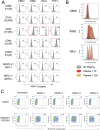
Figure 1
Figure 1
The CEM1 cell line displays a paucity of surface expressed FcRs, a corresponding low level of IgG binding from human plasma, and when transfected to express human DC-SIGN (CD109), is readily infectable with all four serotypes of DENV. (A) CEM1, K562, RAJI and THP-1 cells were assessed for surface expression using monoclonal antibodies specific for FcαR (CD89), FcγRI (CD64), FcγRII (CD32), FcγRIII (CD16) and FcμR (FAIM3). K562, RAJI and THP-1 cells express high levels of CD32, while THP-1 cells also express CD64. In contrast, CEM1 cells express none of the common FcRs on their surface. (B) As predicted by the relative expression levels of CD32, RAJI (DMFI at 1:10 = 1851) and K562 (DMFI at 1:10 = 909) cells display substantial levels of IgG binding when incubated with healthy human donor plasma, whereas CEM1 (DMFI at 1:10 = 71) cells show a correspondingly low level of IgG binding from healthy human donor plasma. (C) The CEM2001 cell line (DC-SIGN transfected CEM1 cells; see Supplementary Figure 1 ) is permissive to infection by all four serotypes of DENV. Percent infection was assessed 24 hours after incubation with 5 PFU per cell of each serotype.
Figure 2
Figure 2
CEM2001 cells were infected for 2 hours with 50 PFU per cell of each of the serotypes of DENV and then treated, or not, with Trypsin/EDTA and incubated for a further 24 hours. (A) Surface antigen expression was assessed using DENV-immune plasma and a secondary goat anti-human IgG antibody. A clear increase in de novo surface antigen expression occurs for all four serotypes from 2 hours post-infection to 24 hours post-infection. No antibody-binding (opsonization) occurs when DENV-naïve plasma is used. (B) The gating strategy used for DENV-4 is depicted. Some binding of virions (from the inoculum) directly to the surface of the DC-SIGN positive cell line is evident at 2 hours, however trypsin treatment clearly removes these virions. The subsequent increase in antigen expression at 24 hours is quantified as the DMFI. Supplementary Figure 2 shows the equivalent gating strategy and data for DENV-1, DENV-2 and DENV-3.
Figure 3
Figure 3
Opsonizing antibody binding is restricted to DENV-infected cells. CEM2001 were infected with serial dilutions of DENV to reach approximately 50% of cells infected, then stained for intracellular 2H2 expression (marker of infection), and surface IgG binding from DENV-immune plasma. Columns (A–E) progressively show the gating strategy for light scatter, identification of infected cells and an overlay of cell infection versus IgG opsonization with DENV-immune plasma. For each of the four serotypes of DENV, infected cells (2H2-positive) were shaded in red while uninfected are shaded black. There is a clear preferential binding of IgG from DENV-immune plasma to infected cells versus uninfected cells. The increase in IgG binding is quantified as the DMFI. Supplementary Figure 3 shows a complete outline of the gating strategy for each serotype with uninfected cultures and DENV-naïve plasma included as controls.
Figure 4
Figure 4
Viral antigen expression remains constant as the force of infection varies. CEM2001 cells were infected with titrated amounts (PFU per cell) of each DENV serotype and stained for intracellular 2H2 expression and surface NS1 (7E11 MAb) and surface E expression (4G2 MAb). (A, B) Intracellular 2H2 expression was assessed at 24 hours post-infection with each of the four serotypes of DENV (5 PFU per cell). Uninfected control cells were overlayed for each infection culture and presented as histograms. 2H2 negative cells clearly overlap with the control cell peak. (C) Surface expression of E and NS1 antigen was assessed for different levels of viral infection of target cells. As the force of infection deceases (percent infection) the MFI of intracellular 2H2 remains the same for each serotype, while surface expression of E (4G2) and NS1 (7E11) remains constant per cell. (D) The gating strategy for detecting surface expression of DENV E protein and NS1 glycoprotein is shown for all four serotypes. Control staining, secondary goat anti-mouse BV421 staining, as well as 4G2 and 7E11 are shown in dot-plots against intracellular 2H2 (directly conjugated 2H2-AF488).
Figure 5
Figure 5
DENV can be maintained in CEM2001 cells in long-term cultures using an uninfected cell replenishment strategy. (A) CEM2001 cells were infected with all four serotypes of DENV and then maintained in continuous culture for 45 days. The level of infection was determined using intracellular 2H2 staining. (B) The continuous cultures above were split at day 18 and replenished with uninfected CEM2001 at days indicated by an asterisk* (days 18, 25, 35 and 45). An add back ratio of 4:1 uninfected cell to infection culture cell ratio was used.
Figure 6
Figure 6
DENV can be maintained in CEM2001 cells through 10 passages of weekly replenishment and retains remarkably similar cell and virus replication kinetics. Infected cells from day 42 of the replenishment infection cultures ( Figure 5B ) were considered “passage 1” with virus adapted to growth in the CEM cell background. Nine further passages of weekly replenishment were performed for each serotype with the replenishment ratio denoted. The following parameters were assessed at initiation, day 3 and day 7 of each passage: (A) Fold increase in total viable cells for each serotype; (B) Percent infected cells for each serotype; (C) Fold increase in infected cell number for each serotype.
Figure 7
Figure 7
Anti-DC-SIGN MAb viral inhibition curves for all four serotypes on CEM2001 cells (A–D for DENV-serotypes 1–4 respectively). Percent inhibition for each of the noted MAbs is shown and was measured at 3 days after culture initiation. These curves were used to calculate the IC50 values presented in Table 1 . Test MAbs were 9A9E8, DCS-8C1 and A20120B, while control MAbs included isotype matched antibodies and two MAbs that bind surface proteins on CEM2001 but should not be involved in viral entry (anti-CD4 and anti-CD317).
Figure 8
Figure 8
A panel of 67 previously characterized MAbs against DENV were tested for binding to DENV-infected cells. All MAbs were synthesized with a human IgG1 Fc region as previously described (32). Data is shown for infected cells (2H2 positive) for all serotypes (A–D) and is presented as the ΔMFI compared to a secondary antibody only control (goat anti-human IgG conjugated to Alexafluor 647). The gating strategy and examples of positive and negative binding MAbs is shown in Supplementary Figure 8 .
Figure 9
Figure 9
DENV-infected cells are applicable target cells for ADCP assays. (A) Plasma (dilued 1:100) from two DENV-naïve subjects (ID15; ID66) and two DENV-immune subjects (SC2; SC4) were compared for ADCP capacity using PKH26 dye staining followed by co-incubation with PBMC-derived CD14+ monocytes. Controls include all cells with no antibody source and uninfected CEM2001 cells. A clear signal (s/n = 5.3× to 8.5×) is seen for all four serotypes using DENV-immune plasma. (B, C) Titration of the ADCP response was conducted for DENV-plasma and data presented the delta percent PKH26+/CD14+ monocytes compared with no antibody addition. The gating strategy for CD14+ monocytes is shown in Supplementary Figure 9 .
Figure 10
Figure 10
DENV-infected cells (2H2+) are suitable target cells for ADCD assays. Plasma (diluted 1:25) from two DENV-naïve subjects (ID15; ID66) and two DENV-immune subjects (SC2; SC4) were compared for ADCD activity using plasma antibody-opsonized infected target cells co-incubated with Low-Tox New Guinea Pig complement. C3 deposition was detected using FITC-conjugated goat anti–Guinea Pig Complement C3 antibody and analyzed by flow cytometry. Controls include all cells with no antibody source and uninfected CEM2001 cells. The gating strategy for DENV-infected target cells showing C3 deposition and ADCD activity using DENV-immune plasma is shown in Supplementary Figure 10A . Comparative analysis of ADCD activity on DENV-uninfected cells (2H2-) from the same plasma and same experiment is shown in Supplementary Figure 10B .

References

    1. Guzman MG, Vazquez S. The complexity of antibody-dependent enhancement of dengue virus infection. Viruses. (2010) 2:2649–62. doi: 10.3390/v2122649 - DOI - PMC - PubMed
    1. Bhatt S, Gething PW, Brady OJ, Messina JP, Farlow AW, Moyes CL, et al. The global distribution and burden of dengue. Nature. (2013) 496:504–7. doi: 10.1038/nature12060 - DOI - PMC - PubMed
    1. Guzman MG, Harris E. Dengue. Lancet. (2015) 385:453–65. doi: 10.1016/S0140-6736(14)60572-9 - DOI - PubMed
    1. Improving data for dengue. Available online at: https://www.who.int/activities/improving-data-for-dengue (Accessed July 30, 2024).
    1. Messina JP, Brady OJ, Golding N, Kraemer MUG, Wint GRW, Ray SE, et al. The current and future global distribution and population at risk of dengue. Nat Microbiol. (2019) 4:1508–15. doi: 10.1038/s41564-019-0476-8 - DOI - PMC - PubMed

LinkOut - more resources