[Preprint]. 2025 Jul 3:2025.07.02.662830.
doi: 10.1101/2025.07.02.662830.

PCNA is a Nucleotide Exchange Factor for the Clamp Loader ATPase Complex

Affiliations

PCNA is a Nucleotide Exchange Factor for the Clamp Loader ATPase Complex

Joshua Pajak et al. bioRxiv. .

Abstract

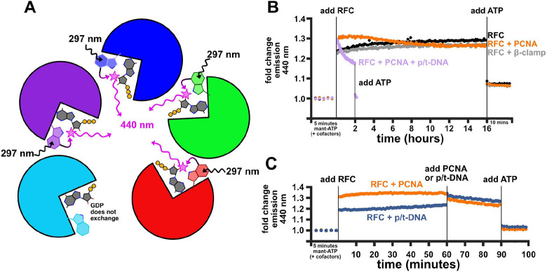
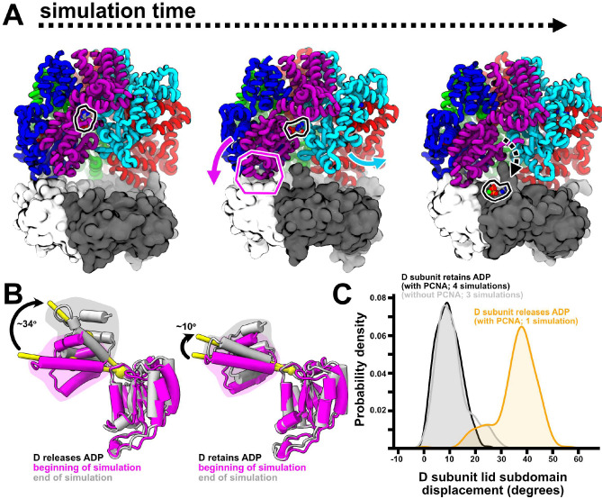
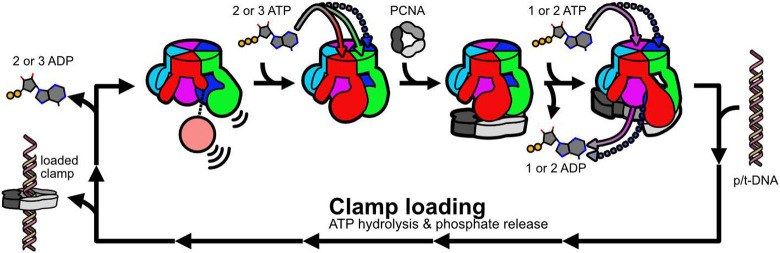
All life requires loading ring-shaped sliding clamp protein complexes onto DNA. The sliding clamp loader is a conserved AAA+ ATPase that binds the sliding clamp, opens the ring, and places it onto DNA. While recent structural work on both the canonical and 'alternative' clamp loaders has shed light into how these machines perform their task once, it remains unclear how clamp loaders are recycled to load multiple sliding clamps. Here, we present structures of the Saccharomyces cerevisiae clamp loader Replication Factor C (RFC) in absence of sliding clamp or supplemented nucleotide. Our structures indicate that RFC holds onto ADP tightly in at least two of its four ATPase active sites, suggesting that nucleotide exchange is regulated. Our molecular dynamics simulations and biochemical data indicate that binding of the sliding clamp PCNA causes rapid exchange of tightly bound ADP. Our data suggests that PCNA acts as a nucleotide exchange factor by prying apart adjacent subunits, providing a pathway for ADP release. We propose that, by using its own substrate as a nucleotide exchange factor, RFC excludes off-pathway states that would arise from binding DNA prior to PCNA.


Figures

Figure 1.
Figure 1.. Cryo-EM reconstructions of the apo yeast RFC complex.
A. Six reconstructions after 3D classification are depicted as viewed from the PCNA-interacting interface, and color coded by local resolution estimation. B. A single reconstruction is highlighted. (left) The reconstruction and model are is color coded by the RFC subunit (right). C. Gaussian surfaces of the model shown in panel B (left) and the autoinhibited RFC:PCNA complex (right; PDB: 7thj; PCNA not shown for clarity) are superimposed (center; apo colorful, autoinhibited transparent) to highlight the missing density corresponding to the AAA+ domain of the A subunit.
Figure 2.
Figure 2.. Cryo-EM reconstructions reveal mixed nucleotide occupancy of the ‘apo’ RFC complex.
A. All six models are superimposed on the D subunit. The C subunit is silver if it is apo and blue if it is ADP-bound. Sub-panels show the Walker A motif and bound nucleotide from a representative class. B. Centers-of-mass of the Rossmann domains of the A-D subunits are shown as yellow spheres and superimposed on Gaussian surfaces of modeled RFC. Two models from this work are shown, as well as the autoinhibited RFC:PCNA complex (PDB: 7thj; PCNA not shown for clarity) and the crab-claw open RFC:PCNA complex (PDB: 7ti8; PCNA not shown for clarity).
Figure 3.
Figure 3.. Nucleotide-dependent conformational changes of RFC subunits.
A. Nucleotide-bound RFC subunits (ATP-γS-bound from the autoinhibited RFC:PCNA complex or ADP-bound from this work; silver) and apo subunits (colorful) are aligned on their ATPase active sites. The centers-of-mass of lid subdomains and the pivot point residues are shown as yellow spheres, and the angular displacement of the centers-of-mass is reported. The lid subdomain is highlighted by a shaded region. B. ADP-bound (colorful) and ATP-γS-bound (silver; from autoinhibited RFC:PCNA complex) subunits are aligned on their ATPase active sites. The centers-of-mass of lid subdomains and the pivot points are shown as yellow spheres, and the angular displacement of the centers-of-mass is shown. The lid subdomain is highlighted by a shaded region. C. Apo (colorful) and nucleotide-bound (silver) active sites are aligned. The N-terminal loop is highlighted by a shaded region.
Figure 4.
Figure 4.. Molecular dynamics simulations of RFC and predicted relative ADP dissociation rates.
A. The sampled distances between centers-of-mass of Rossmann domains from ATP-bound (black) and ADP-bound (orange) equilibrium molecular dynamics simulations are shown as kernel density estimates. The distances from the structures reported in Fig. 2B are shown as dashed lines for reference. B. Results from τ-RAMD simulations. (left) ADP residence probability is plotted against simulation time, and ADP residence time from each simulation is plotted on the right. Solid bars indicate the half-survival time, and red dots indicate right-censored data (where the unbinding event was not sampled during the simulation after one day of wall-time).
Figure 5.
Figure 5.. MANT-ATP FRET assays show that PCNA promotes fast nucleotide exchange in RFC.
A. A cartoon schematic of the assay. Tryptophan residues are excited at 297 nm and can transfer energy if MANT-labeled ATP is bound, which emits at 440 nm. Because the E subunit is always GDP-bound, we assume that it does not exchange and contribute to signal in this assay. B. The fold change of measured emission at 440 nm is reported for different conditions. In all cases, the average intensity from the five minutes of measurement prior to adding RFC was used to set the baseline. C. Sequentially adding RFC’s binding partners shows that PCNA and not p/t-DNA is the primary driver of nucleotide exchange.
Figure 6.
Figure 6.. MD simulations predict how PCNA promotes ADP release.
A. Snapshots from an equilibrium simulation show that as the ADP-bound D subunit engages with PCNA, it is pried away from the E subunit and ADP is released from its active site. B. ADP release from the D subunit coincides with a large rotation of the lid subdomain in the same direction observed in the experimentally determined structure of the C subunit (compare with Fig. 3A). C. Sampled distribution of the D subunit’s lid subdomain rotation from equilibrium simulations of ADP-bound RFC without PCNA (silver), RFC:PCNA where ADP is not released (black), and RFC:PCNA where ADP is released (orange) is shown as kernel density estimates.
Figure 7.
Figure 7.. A speculative model for the role of nucleotide exchange in RFC.
Mixed nucleotide occupancy RFC has flexible apo subunits (A and B) and more rigid ADP-bound subunits (C and D). Two or three ATP molecules binding at A, B, and possibly C stabilizes RFC in a conformation competent to bind PCNA. PCNA binding promotes additional ADP release from the remaining subunits, and ATP binding drives crab-claw opening and downstream clamp loading.

Similar articles

References

    1. Kelch B. A., The lord of the rings: Structure and mechanism of the sliding clamp loader. Biopolymers 105, 532–546 (2016). - PubMed
    1. Choe K. N., Moldovan G.-L., Forging ahead through darkness: PCNA, still the principal conductor at the replication fork. Molecular cell 65, 380–392 (2017). - PMC - PubMed
    1. Moldovan G.-L., Pfander B., Jentsch S., PCNA, the maestro of the replication fork. Cell 129, 665–679 (2007). - PubMed
    1. Erzberger J. P., Berger J. M., Evolutionary relationships and structural mechanisms of AAA+ proteins. Annu. Rev. Biophys. Biomol. Struct. 35, 93–114 (2006). - PubMed
    1. Davey M. J., Jeruzalmi D., Kuriyan J., O’Donnell M., Motors and switches: AAA+ machines within the replisome. Nat Rev Mol Cell Biol 3, 826–835 (2002). - PubMed

Publication types

LinkOut - more resources