[Preprint]. 2025 Jun 30:2025.06.27.661568.
doi: 10.1101/2025.06.27.661568.

SARS-CoV-2 infection disrupts syncytial and endothelial integrity and alters PLGF levels in the placenta

Affiliations

SARS-CoV-2 infection disrupts syncytial and endothelial integrity and alters PLGF levels in the placenta

Brittany R Jones et al. bioRxiv. .

Update in

Abstract

Introduction: SARS-CoV-2 infection during pregnancy has been associated with an increased risk for several pregnancy-related disorders, particularly preeclampsia (PE). However, there are limited studies determining the impact of SARS-CoV-2 on placental physiology and function.

Methods: Placental samples were acquired from two large prospective cohorts: STOP-COVID19 and REBRACO studies. Placental villous tissues (VTs) were collected from pregnant women who tested positive for SARS-CoV-2 without PE during pregnancy. Immunohistochemistry and immunofluorescence were used to assess pathological features known to be altered in PE, including 1) syncytial knot formation; 2) alterations in renin-angiotensin system components; 3) and endothelial integrity. Maternal serum was collected to examine AT1 autoantibodies levels using an immunoassay.

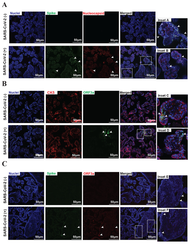
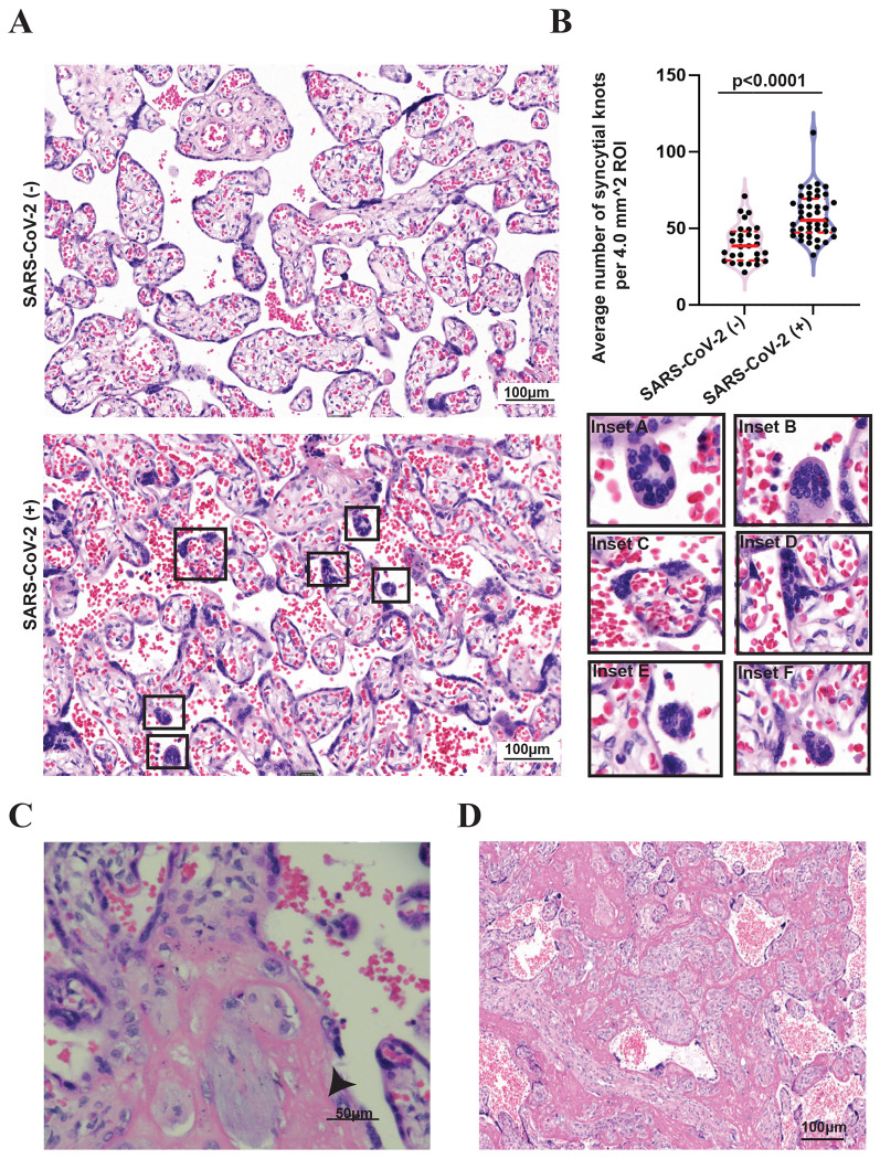
Results: SARS-CoV-2 viral proteins spike, nucleocapsid, and ORF3a were observed in the syncytiotrophoblast layer and stroma of placental VT. SARS-CoV-2-infected placentas exhibited increased numbers of syncytial knots, which were positive for Flt-1 and SARS-CoV-2 viral proteins. In addition, the presence of placental infarctions and excessive fibrin deposits was also observed in infected placentas. Infection was associated with decreased placental expression of PlGF and an increase in the placental Flt-1/PlGF expression ratio, mostly driven by PlGF. No significant changes in maternal serum AT1AA levels were observed. Finally, SARS-CoV-2-infected placentas exhibited a significant decrease in vimentin expression.

Discussion: SARS-CoV-2 infection negatively impacts placental integrity in the form of increased syncytial knots, dysregulated RAS components, and endothelial damage. Since all these features are similarly disrupted in PE, this could be a mechanism through which SARS-CoV-2 infection during pregnancy increases the risk of a PE-like syndrome.

Keywords: COVID-19; Preeclampsia; Renin Angiotensin system; Syncytial knots, placenta.


Conflict of interest statement

Declaration of interest IUM serves on the scientific advisory board of Seed Health. The authors declare that they have no known competing financial interests or personal relationships that could have appeared to influence the work reported in this paper.

Figures

Figure 1:
Figure 1:. Foci of SARS-CoV-2 infection in the placenta.
(A) Detection of SARS-CoV-2 proteins spike (green), nucleocapsid (red), and colocalization (inset A and B) in villous tissue from SARS-CoV-2 positive term placentas (magnification scale 20X; scale bar represents 50 μm), Nuclei (blue). (B) Presence of SARS-CoV-2 ORF3a (green) in STB layer (Inset C and D); STB layer outlined with CK5 (red) (magnification scale 20X; scale bar represents 50 μm). (C) SARS-CoV-2 proteins spike (green) and ORF3a (red) detected in villous tissue and colocalization of (Inset E and F) of viral proteins in STB layer (magnification scale 20X; scale bar represents 50 μm).
Figure 2:
Figure 2:. Increased formation of syncytial knots in SARS-CoV-2 infected placentas.
(A) Presence of SK in SARS-CoV-2 positive placentas. Syncytial knots were located in the maternal circulation (Inset A, C, and D) or suspended between villi (Inset B, E, and F). (B) Semiquantitive analysis of syncytial knots formation in uninfected and SARS-CoV-2 infected placentas. Values are expressed as mean +/− standard error of the mean (SEM); * p <0.05, ** p < 0.01, ***: p < 0.001; ns: non-significant. (C-D) Presence of placental infarction (C), and excessive fibrin deposition (D) in SARS-CoV-2 infected placentas.
Figure 3:
Figure 3:. SARS-CoV-2 infection is associated with changes in RAS components in the placenta.
(A) Immunofluorescence images of uninfected and SARS-CoV-2 infected villous tissue showing expression of PlGF (red), Flt-1 (green) (magnification scale 20X; scale bar represents 100 μm). Presence of Flt-1 in SK of SARS-CoV-2-infected placentas (Inset A and B). (B-C) Quantification of mean fluorescence intensity of PlGF and Flt-1 in uninfected and infected placentas (B) and Flt-1/PlGF ratio (C). (D) Pre-delivery of angiotensin II type 1-receptor autoantibody (AT1-AA) levels in sera of uninfected and SARS-CoV-2-infected pregnant women. Bars represent means and error bars represent standard error of the mean (SEM); *: * p <0.05, ** p < 0.01, ***: p < 0.001; ns: nonsignificant difference.
Figure 4:
Figure 4:. SARS-CoV-2 infection impacts endothelial integrity of the placenta.
(A) Foci of chorionic villi with more than ten capillaries in more than ten villi, under objective lens of 10x magnification, characterizing chorangiosis (scale bar represents 100 μm). (B) Expression of vimentin (red) in villous tissue of uninfected (Inset A and B) and infected placentas (Inset C and D) (magnification scale 20X; scale bar represents 100μm). (C) Quantification of mean fluorescence intensity of vimentin in uninfected and SAR-CoV-2 infected placentas, bars represent means and error bars represent standard error of the mean (SEM); *: * p <0.05, ** p < 0.01, ***: p < 0.001; ns: nonsignificant difference.

References

    1. Kumar D., Verma S., Mysorekar I.U., COVID-19 and pregnancy: clinical outcomes; mechanisms, and vaccine efficacy, Translational Research 251 (2023) 84–95. 10.1016/j.trsl.2022.08.007. - DOI - PMC - PubMed
    1. Sathiya R., Rajendran J., Journal S.S.-M.M., undefined 2022, COVID-19 and Preeclampsia: Overlapping Features in Pregnancy, Ncbi.Nlm.Nih.Gov (n.d.). https://www.ncbi.nlm.nih.gov/pmc/articles/PMC8798587/ (accessed December 6, 2022). - PMC - PubMed
    1. Boettcher L.B., Metz T.D., Maternal and neonatal outcomes following SARS-CoV-2 infection, Semin Fetal Neonatal Med 28 (2023) 101428. 10.1016/j.siny.2023.101428. - DOI - PMC - PubMed
    1. Wong C.K.H., Lau K.T.K., Chung M.S.H., Au I.C.H., Cheung K.W., Lau E.H.Y., Daoud Y., Cowling B.J., Leung G.M., Nirmatrelvir/ritonavir use in pregnant women with SARS-CoV-2 Omicron infection: a target trial emulation, Nat Med 30 (2024) 112–116. 10.1038/s41591-023-02674-0. - DOI - PubMed
    1. Maher M., Owens L., SARS-CoV-2 infection and female reproductive health: A narrative review, Best Pract Res Clin Endocrinol Metab 37 (2023) 101760. 10.1016/j.beem.2023.101760. - DOI - PMC - PubMed

Publication types