Review
. 2025 Aug;137(2):e70074.
doi: 10.1111/bcpt.70074.

Coronary Microvascular Dysfunction in Ischaemic Heart Disease: Lessons From Large Animal Models

Affiliations
Review

Coronary Microvascular Dysfunction in Ischaemic Heart Disease: Lessons From Large Animal Models

Oana Sorop et al. Basic Clin Pharmacol Toxicol. 2025 Aug.

Abstract

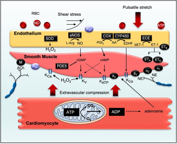
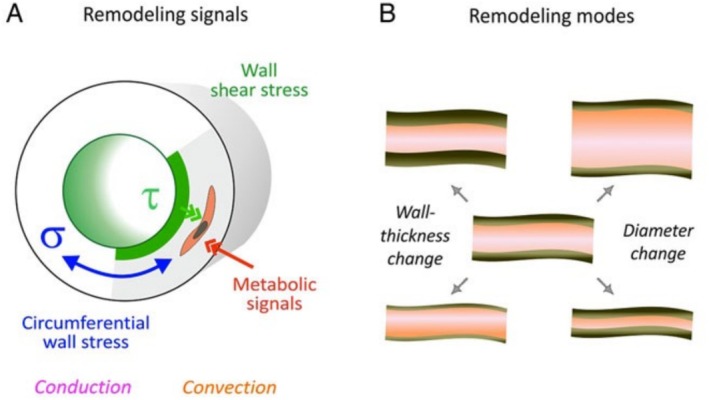
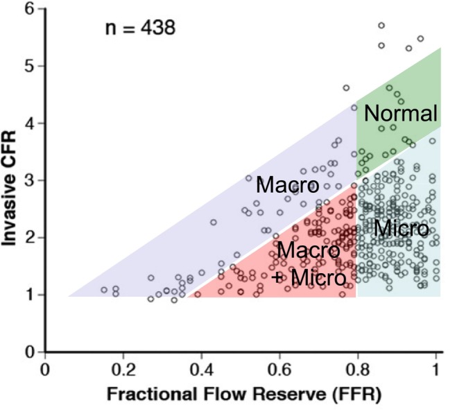
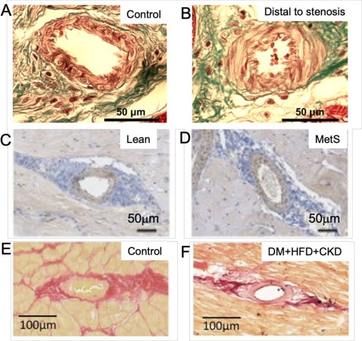
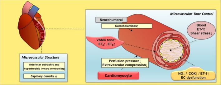
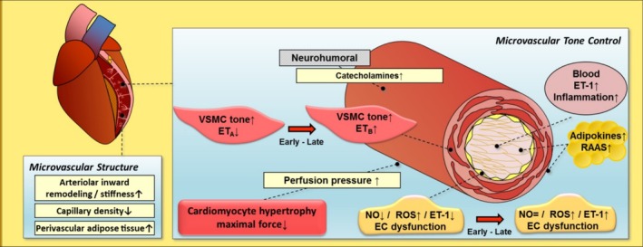
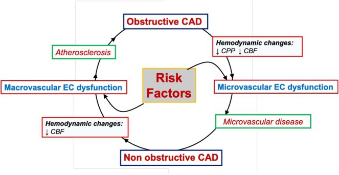
The coronary microvasculature is principally responsible for matching coronary blood flow to myocardial demand of oxygen and nutrients. Short-term control of coronary blood flow is achieved via alterations in coronary microvascular tone, whereas long-term control of coronary flow also involves remodelling of the coronary microvasculature, including adjustments in vascular structure, diameter and density. In the past 50 years, considerable research efforts have been directed at understanding the functional and structural coronary microvascular adaptations involved in matching myocardial oxygen supply to demand, and how these mechanisms are affected by various diseases. In this review article, we will discuss our current understanding of the mechanisms underlying the regulation of coronary microvascular tone under healthy physiological conditions and in ischaemic heart disease. We will specifically discuss the role of microvascular dysfunction in obstructive and non-obstructive coronary artery disease, as studied in large animal models and confirmed in human studies. Future research should be directed at further unravelling the disease-specific mechanisms of coronary microvascular dysfunction in order to identify therapeutic targets to improve microvascular function in patients with ischaemic heart disease.

Keywords: INOCA; coronary artery disease; coronary blood flow; endothelial dysfunction; ischaemic heart disease; microvascular dysfunction.


Conflict of interest statement

The authors declare no conflicts of interest.

Figures

FIGURE 1
FIGURE 1
Schematic drawing of endothelium, vascular smooth muscle cell (VSMC) and cardiomyocyte illustrating mechanisms for control of vasomotor tone and diameter. A2, adenosine receptor 2; AA, arachidonic acid; ACh, acetylcholine; ADP, adenosine diphosphate; ATP, adenosine triphosphate; bET‐1, big endothelin‐1; CO2, carbon dioxide; COX, cyclooxygenase; CYP2C9, cytochrome P450 2C9; ECE, endothelin‐converting enzyme; EETs, epoxyeicosatrienoic acids; eNOS, endothelial nitric oxide synthase; ETA, endothelin type A receptor; ETB, endothelin type B receptor; ET‐1, endothelin‐1; H2O2, hydrogen peroxide; KATP, ATP‐sensitive K+ channel; KCa, calcium‐activated K+ channel; KV, voltage‐gated K+ channel; L‐arg, L‐arginine; M, muscarinic receptor; NE, norepinephrine; NO, nitric oxide; O2 oxygen; O2 , superoxide anion; P2, purinergic receptor type 2; P2y, purinergic receptor type 2y; PDE5, phosphodiesterase; PGI2, prostacyclin; SOD, superoxide dismutase; α1, α1‐adrenergic receptor; α2, α2‐adrenergic receptor; β2, β2‐adrenergic receptor. Adapted with permission from Sorop et al., Microcirculation, M. Dorobantu and L. Badimon (eds.) Springer 2020.
FIGURE 2
FIGURE 2
Mechanisms of vascular remodelling: (A) endothelial wall shear (τ), circumferential wall stress (σ) and metabolic signals may act as vasoconstrictor or vasodilator stimuli resulting in changes in vascular diameter and wall mass (B). With permission from Pries et al. [23].
FIGURE 3
FIGURE 3
Invasive measurements of individual coronary flow reserve (CFR) and fractional flow reserve (FFR) values in patients with suspected ischaemic heart disease. An FFR value below 0.8 identifies patients with a haemodynamically significant coronary artery stenosis, either without (Macrovascular, Macro) or with microvascular abnormalities (Macro + Micro), while the majority of the patients (blue) showed decreased values of CFR despite a normal FFR, indicative of principally microvascular disease (Micro). Adapted with permission from [50].
FIGURE 4
FIGURE 4
Typical examples of porcine coronary small arteries from healthy myocardium and from myocardium distal to a critical coronary artery stenosis (A, B, trychrome [51]), from lean and metabolic syndrome (MetS) Ossabaw swine (C, D, immunostaining for receptor for advanced glycation end products [52]), and from healthy swine and swine with diabetes mellitus (DM), high‐fat diet (HFD) and chronic kidney disease (CKD) (E, F, picrosirius red [53]). Adapted with permission.
FIGURE 5
FIGURE 5
Microvascular dysfunction distal to a chronic coronary artery stenosis. COX, cyclooxygenase; EC, endothelial cell; ETA, endothelin receptor A; ETB, endothelin receptor B; ET‐1, endothelin‐1; NO, nitric oxide; VSMC, vascular smooth muscle cell. Adapted with permission from Sorop et al., Microcirculation, M. Dorobantu and L. Badimon (eds.) Springer 2020.
FIGURE 6
FIGURE 6
Microvascular dysfunction in the presence of metabolic dysregulation. EC, endothelial cell; ETA, endothelin receptor A; ETB, endothelin receptor B; ET‐1, endothelin‐1; NO, nitric oxide; RAAS, renin angiotensin aldosterone system; ROS, reactive oxygen species; VSMC, vascular smooth muscle cell. Adapted with permission from Sorop et al., Microcirculation, M. Dorobantu and L. Badimon (eds.) Springer 2020.
FIGURE 7
FIGURE 7
Interplay between coronary microvascular and macrovascular dysfunction in ischaemic heart disease. Exposure to cardiovascular risk factors induces early coronary microvascular endothelial dysfunction in conjunction with arterial (inward) remodelling and reduced capillary densities, contributing to myocardial ischaemia. Subsequently, impaired coronary flow may promote macrovascular endothelial dysfunction and development of a proximal coronary artery occlusion, leading to further perfusion impediments and exacerbating microvascular dysfunction and remodelling.

References

    1. Feigl E. O., “Coronary Physiology,” Physiological Reviews 63, no. 1 (1983): 1–205. - PubMed
    1. Tune J. D., Gorman M. W., and Feigl E. O., “Matching Coronary Blood Flow to Myocardial Oxygen Consumption,” Journal of Applied Physiology 97, no. 1 (2004): 404–415. - PubMed
    1. Duncker D. J. and Bache R. J., “Regulation of Coronary Blood Flow During Exercise,” Physiological Reviews 88, no. 3 (2008): 1009–1086, 10.1152/physrev.00045.2006. - DOI - PubMed
    1. Goodwill A. G., Dick G. M., Kiel A. M., and Tune J. D., “Regulation of Coronary Blood Flow,” Comprehensive Physiology 7, no. 2 (2017): 321–382, 10.1002/cphy.c160016. - DOI - PMC - PubMed
    1. Chilian W. M., Eastham C. L., and Marcus M. L., “Microvascular Distribution of Coronary Vascular Resistance in Beating Left Ventricle,” American Journal of Physiology 251, no. 4 (1986): H779–H788. - PubMed

MeSH terms

LinkOut - more resources