Multicenter Study
. 2025 Oct 22;46(40):4090-4101.
doi: 10.1093/eurheartj/ehaf387.

Cardiac amyloidosis detection from a single echocardiographic video clip: a novel artificial intelligence-based screening tool

Affiliations
Multicenter Study

Cardiac amyloidosis detection from a single echocardiographic video clip: a novel artificial intelligence-based screening tool

Jeremy A Slivnick et al. Eur Heart J. .

Abstract

Background and aims: Accurate differentiation of cardiac amyloidosis (CA) from phenotypic mimics remains challenging using current clinical and echocardiographic techniques. The accuracy of a novel artificial intelligence (AI) screening algorithm for echocardiography-based CA detection was assessed.

Methods: Utilizing a multisite, multiethnic dataset (n = 2612, 52% CA), a convolutional neural network was trained to differentiate CA from phenotypic controls using transthoracic apical four-chamber video clips. External validation was conducted globally across 18 sites including 597 CA cases and 2122 controls. Classification accuracy was assessed on the entire external validation dataset, and subgroup analyses were performed both on technetium pyrophosphate scintigraphy referrals, and individuals matched for age, sex, and wall thickness. Model accuracy was also compared with the transthyretin CA score and the increased wall thickness score within a subset of older heart failure with preserved ejection fraction patients with increased wall thickness.

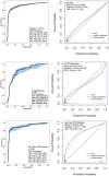
Results: Cardiac amyloidosis patients and controls displayed similar age, sex, race, and comorbidities. After the removal of uncertain AI predictions (13%), model discrimination and classification were excellent for the entire external validation dataset [area under the receiver operating characteristic curve (AUROC) 0.93, sensitivity 85%, specificity 93%], irrespective of CA subtype (sensitivity: light-chain = 84%, wild-type transthyretin = 85%, and hereditary transthyretin = 86%). Performance was maintained in subgroup analysis in patients clinically referred for technetium pyrophosphate scintigraphy imaging (AUROC 0.86, sensitivity 77%, specificity 86%) and matched patients (AUROC 0.92, sensitivity 84%, specificity 91%). The AI model (AUROC 0.93) also outperformed transthyretin CA score (AUROC 0.73) and increased wall thickness (AUROC 0.80) scores.

Conclusions: This AI screening model-using only an apical four-chamber view-effectively differentiated CA from other causes of increased left ventricular wall thickness.

Keywords: Amyloid; Artificial intelligence; Cardiomyopathy; Echocardiography; Heart failure.


Figures

Structured Graphical Abstract
Geographic representation of the separate training, tuning, and international, multi-ethnic external validation cohorts which included patients with cardiac amyloidosis (CA) and controls referred for transthoracic echocardiography. The artificial intelligence model, based on a single apical four-chamber echocardiographic videoclip, was validated in the entire external validation cohort of 2719 patients, in which prevalence of CA was 22%, with AUC 0.93. The model's accuracy was maintained in testing in various subgroups.
Figure 1
Figure 1
Discrimination, classification and calibration of the artificial intelligence model in independent testing datasets. Left-hand side: Receiver operating characteristic curves are depicted with 95% confidence intervals shaded. Statistics are presented on the dataset characteristics, including artificial intelligence discrimination and classification performance. Right-hand side: Smoothed flexible calibration curves present the actual and artificial intelligence-predicted probability of disease. Histograms at the bottom of each plot indicate the distribution of artificial intelligence-predicted probabilities. Both logistic and non-parametric flexible calibration curves are presented to visualize the effect of the underlying data distribution. AUC, area under the curve; NPV, negative predictive value; PPV, positive predictive value; Sens, sensitivity; Spec, specificity; Tc-PYP, technetium pyrophosphate scintigraphy
Figure 2
Figure 2
Discrimination and classification of the artificial intelligence model, transthyretin cardiac amyloidosis score, and increased wall thickness score. (A) Receiver operating characteristic curves for the artificial intelligence model, transthyretin cardiac amyloidosis score, and increased wall thickness score. Decision thresholds for the artificial intelligence model, the transthyretin cardiac amyloidosis score, and the increased wall thickness score were 0.06, 6, and 8, respectively. The reported statistics and confidence intervals represent the median, 2.5th and 97.5th percentiles from bootstrapping. (B) Positive predictive value of the artificial intelligence model, transthyretin cardiac amyloidosis score, and increased wall thickness score according to modelled disease prevalence. AI, artificial intelligence; AUC, area under the curve; IWT, increased wall thickness; NPV, negative predictive value; PPV, positive predictive value; Sens, sensitivity; Spec, specificity; TCAS, transthyretin cardiac amyloidosis score
Figure 3
Figure 3
Decision curve analysis. Decision curve analysis comparing the net proportion of cases referred to technetium pyrophosphate scintigraphy (e.g. standardized net benefit) (left) and the net interventions avoided (right) for the artificial intelligence model (blue), the transthyretin cardiac amyloidosis score, the increased wall thickness score, referring all patients, and referring no patients. Disease prevalence has been modelled at 6.3%, as reported by AbouEzzeddine et al. The modelled clinical decision is whether to refer patients for Tc-PYP imaging, having ruled out positive light chains already, based on the output of the artificial intelligence model or clinical scores. The net proportion of cases referred to technetium pyrophosphate scintigraphy (standardized net benefit, left panel y-axis) reflects the net proportion of all cardiac amyloidosis patients who would be correctly referred with a given method, as a function of the decision threshold probability. Net interventions avoided describe the proportion of patients who would not be sent for unnecessary technetium pyrophosphate scintigraphy imaging using a given method, as a function of decision threshold probability. The decision threshold probability (x-axis) represents a patient’s/clinician's risk tolerance for undergoing technetium pyrophosphate scintigraphy imaging knowing the harms (unnecessary testing), benefits (diagnosing disease and access to treatment), and potential likelihood of a correct referral. Thus, the x-axis reflects the expected likelihood of a true positive referral (e.g. the dashed line at a decision threshold probability of 0.25 = one cardiac amyloidosis case found for every four referrals). Given the significant life-prolonging benefits of available therapies, a threshold of 0.25 was set for numerical reporting of this analysis, meaning that we would tolerate three incorrect technetium pyrophosphate scintigraphy referrals for every true cardiac amyloidosis patient identified by the tests in clinical practice. At this threshold, the artificial intelligence model, increased wall thickness, and transthyretin cardiac amyloidosis score identified a net proportion (e.g. without any false positives) 42.3%, 5.9%, and 0% of cases, indicating a 36.4% benefit of the artificial intelligence model over increased wall thickness. AI, artificial intelligence; IWT, increased wall thickness; TCAS, transthyretin cardiac amyloidosis score; Tc-PYP, technetium pyrophosphate scintigraphy

References

    1. Maurer MS, Schwartz JH, Gundapaneni B, Elliott PM, Merlini G, Waddington-Cruz M, et al. Tafamidis treatment for patients with transthyretin amyloid cardiomyopathy. N Engl J Med 2018;379:1007–16. 10.1056/NEJMoa1805689 - DOI - PubMed
    1. Barrett CD, Dobos K, Liedtke M, Tuzovic M, Haddad F, Kobayashi Y, et al. A changing landscape of mortality for systemic light chain amyloidosis. JACC Heart Fail 2019;7:958–66. 10.1016/j.jchf.2019.07.007 - DOI - PubMed
    1. Writing C, Kittleson MM, Ruberg FL, Ambardekar AV, Brannagan TH, Cheng RK, et al. 2023 ACC expert consensus decision pathway on comprehensive multidisciplinary care for the patient with cardiac amyloidosis: a report of the American College of Cardiology solution set oversight committee. J Am Coll Cardiol 2023;81:1076–126. 10.1016/j.jacc.2022.11.022 - DOI - PubMed
    1. Arbelo E, Protonotarios A, Gimeno JR, Arbustini E, Barriales-Villa R, Basso C, et al. 2023 ESC guidelines for the management of cardiomyopathies. Eur Heart J 2023;44:3503–626. 10.1093/eurheartj/ehad194 - DOI - PubMed
    1. Dorbala S, Ando Y, Bokhari S, Dispenzieri A, Falk RH, Ferrari VA, et al. ASNC/AHA/ASE/EANM/HFSA/ISA/SCMR/SNMMI expert consensus recommendations for multimodality imaging in cardiac amyloidosis: part 1 of 2-evidence base and standardized methods of imaging. Circ Cardiovasc Imaging 2021;14:e000029. 10.1161/HCI.0000000000000029 - DOI - PubMed

Grants and funding