. 2025 Aug 1;6(4):zqaf030.
doi: 10.1093/function/zqaf030.

Sustained Accumulation of Molecular Clock Suppressors Period 1 and Period 2 Promotes C2C12 Myotube Atrophy Through an Autocrine-Mediated Mechanism With Relevance to Androgen Deprivation-Induced Limb Muscle Mass Loss

Affiliations

Sustained Accumulation of Molecular Clock Suppressors Period 1 and Period 2 Promotes C2C12 Myotube Atrophy Through an Autocrine-Mediated Mechanism With Relevance to Androgen Deprivation-Induced Limb Muscle Mass Loss

Grant R Laskin et al. Function (Oxf). .

Abstract

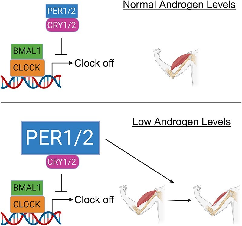
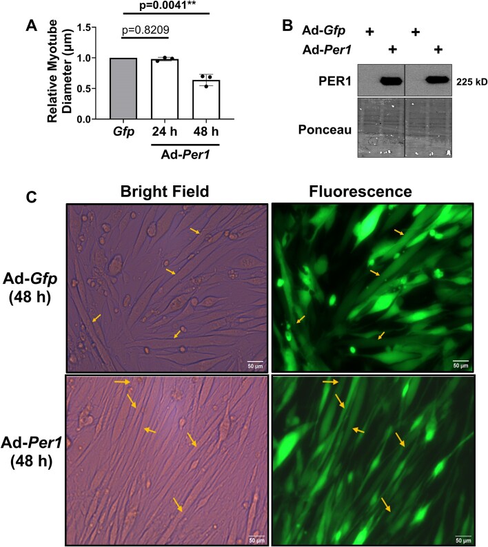
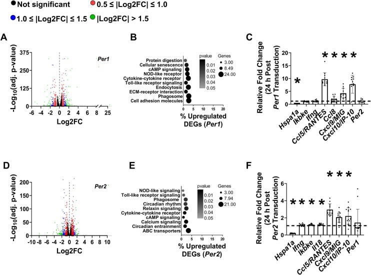
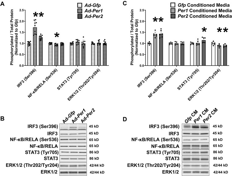
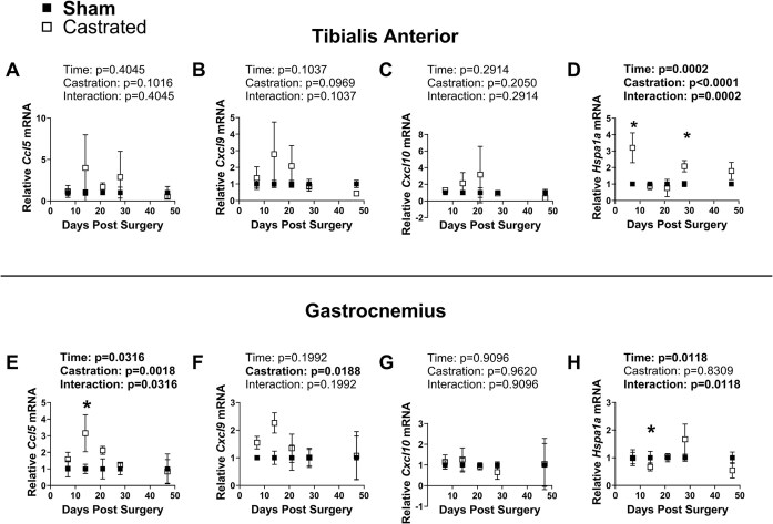
Low testosterone in males (hypogonadism) is associated with limb muscle mass loss, yet the underlying mechanisms of muscle mass loss remain largely unknown. We previously showed androgen deprivation disrupted limb muscle molecular clock function, and the disruption coincided with elevated levels of the primary molecular clock suppressor, Period 2 (Per2). The purposes herein were to determine if PER2 overexpression leads to muscle atrophy and if preventing PER2 accumulation blunts limb muscle mass loss in response to androgen deprivation. Here, we identify Per2 as a negative regulator of muscle size. Overexpression of Per2 in differentiated C2C12 myotubes reduced myotube diameter, while deletion of Per2 in male mice partially preserved tibialis anterior (TA) mass following castration. The muscle-sparing effect of Per2 deletion in vivo was specific to the TA despite evidence of molecular clock disruption and mass loss in other muscles. Subsequently, we show overexpression of the other primary clock suppressor, Period 1 (Per1) also reduced myotube diameter in differentiated C2C12 myotubes. Mechanistically, both Per1 and Per2 overexpression in vitro induced muscle atrophy in part by an autocrine-mediated mechanism likely involving inflammation as their overexpression induced an inflammatory gene expression signature and increased cytokine/chemokine secretion. Moreover, incubation of C2C12 myotubes in the media conditioned from Per1 or Per2 overexpressing myotubes reduced myotube diameter. Several inflammatory genes identified in vitro were also altered in the limb muscles in response to androgen deprivation. These findings identify a previously unrecognized role for Per1/2 in regulating skeletal muscle mass with implications for muscle loss during hypogonadism.

Keywords: RNA sequencing; circadian rhythm; hypogonadism; inflammation.


Conflict of interest statement

The authors declare no conflicts of interest.

Figures

Graphical Abstract
Graphical Abstract
Figure 1.
Figure 1.
Overexpression of Per2 induces muscle atrophy in vitro. (A) The diameters of myotubes overexpressing Per2 relative to the diameters of myotubes overexpressing Gfp were determined at 24 h or 48 h post adenoviral transduction. (B) PER2 protein levels were assessed 24 h post adenoviral transduction by Western blot analysis. (C) Representative images of Gfp and Per2 transduced myotubes 48 h post-transduction. Pictures are at 20×. Scalebar is 50 µm. Arrows indicate myotubes included in the analysis. All C2C12 myotube diameter experiments consisted of 3 independent experimental replicates with each replicate containing separate plates for each virus. The diameters of myotubes from each virus within a replicate were averaged to generate a single diameter value for that replicate. The mean diameter values from each replicate were used in the final analysis.
Figure 2.
Figure 2.
Loss of Per2 preserves some limb muscle mass in response to androgen deprivation. (A-F) % differences in body or organ masses between the castrated group and the corresponding sham group of Per2+/+ and Per2−/− mice 8 wk after a castration or sham surgery. N = 7-10 per group.
Figure 3.
Figure 3.
Overexpression of Per1 induces muscle atrophy in vitro. (A) The diameters of myotubes overexpressing Per1 relative to the diameters of myotubes overexpressing Gfp were determined at 24 h or 48 h post adenoviral transduction. (B) PER1 protein levels were assessed 24 h post adenoviral transduction by Western blot analysis. Line on the blot represents samples run on the same gel but in non-contiguous lanes. (C) Representative images of Gfp and Per1 transduced myotubes 48 h post-transduction. Pictures are at 20×. Scalebar is 50 µm. Arrows indicate myotubes included in the analysis. All C2C12 myotube diameter experiments consisted of 3 independent experimental replicates with each replicate containing separate plates for each virus. The diameters of myotubes from each virus within a replicate were averaged to generate a single diameter value for that replicate. The mean diameter values from each replicate were used in the final analysis.
Figure 4.
Figure 4.
Overexpression of Per1 or Per2 induces an inflammatory gene expression signature in myotubes. (A) Volcano plot illustrating −Log10 fold changes (FC) of genes from C2C12 myotubes overexpressing Per1 relative to myotubes expressing Gfp following RNA sequencing. Labeled dot represents Per1 mRNA data point. (B) Enriched KEGG pathways from upregulated differentially expressed genes (DEGs) from C2C12 myotubes overexpressing Per1 relative to myotubes expressing Gfp. (C) The mRNA contents of heat shock protein 70 (Hspa1a), inhibitor of nuclear factor kappa B kinase subunit epsilon (Ikbke), interferon gamma (Ifng), C-C motif chemokine ligand 5 (Ccl5/RANTES), C-C motif chemokine ligand 8 (Ccl8), C-X-C motif ligand 9 (Cxcl9/MIG), C-X-C motif ligand 10 (Cxcl10/IP-10), and Per2 were measured in myotubes 24 h after transduction with adenovirus overexpressing Per1 and made relative to values in myotubes transduced with adenovirus expressing Gfp. N = 11-13 per group from 3 independent experiments. (D) Volcano plot illustrating −Log10 FC of genes from C2C12 myotubes overexpressing Per2 relative to myotubes expressing Gfp following RNA sequencing. Labeled dot represents Per2 mRNA data point. (E) Enriched KEGG pathways from upregulated DEGs from C2C12 myotubes overexpressing Per2 relative to myotubes expressing Gfp. (C) The mRNA contents of Hspa1a, Ikbke, Ifng, Ccl5/RANTES, Ccl8, Cxcl9/MIG, Cxcl10/IP-10, and Per1 were measured in myotubes 24 h after transduction with adenovirus overexpressing Per2 and made relative to values in myotubes transduced with adenovirus expressing Gfp. N = 11-13 per group from 3 independent experiments. *Significantly different than values in myotubes expressing Gfp at P < 0.05.
Figure 5.
Figure 5.
Per1 or Per2 overexpression induce myotube atrophy via an autocrine-mediated mechanism. (A) Experimental design for conditioned media experiment. (B) Diameter of C2C12 myotubes incubated for 24 h in media conditioned from C2C12 myotubes transduced with adenovirus overexpressing Per1, Per2, or Gfp. (C) Concentrations of C-X-C motif ligand 9 (CCL9/MIG), C-X-C motif ligand 10 (CXCL10/IP-10), Interferon Gamma (IFNγ), CCL5/RANTES, and IL18 in the media conditioned from C2C12 myotubes transduced with adenovirus overexpressing Per1, Per2, or Gfp. N = 5 per transduction condition. *Significantly different than Gfp conditioned media at P < 0.05.
Figure 6.
Figure 6.
Per1 and Per2 overexpression leads to increased phosphorylation of the Interferon Regulated Factor 3 (IRF3) transcription factor. (A) The phosphorylated to total protein ratios for IRF3 (Ser396), Nuclear factor kappa beta/RELA (NF-κB/RELA; Ser536), Signal transducer and activator of transcription 3 (STAT3; Tyr705), and Extracellular signal regulated kinase1/2 (ERK1/2; Thr202/Tyr204) in C2C12 myotubes 24 h after transduction with adenovirus overexpressing Per1, Per2, or Gfp. N = 9 per transduction condition from 3 independent experiments. (B) Representative Western blot. (C) The phosphorylated to total protein ratios for IRF3 (Ser396), NF-κB/RELA (Ser536), STAT3 (Tyr705), and ERK1/2 (Thr202/Tyr204) in C2C12 myotubes 1 h after exposure to media conditioned from C2C12 myotubes transduced with adenovirus overexpressing Per1, Per2, or Gfp for 48 h. N = 6 per condition from 3 independent experiments. (D) Representative Western blot. *Significantly different than either Ad-Gfp or Gfp CM at P < 0.05.
Figure 7.
Figure 7.
Time course of tissue characteristics following castration. (A) body mass, (B) seminal vesicle (SV) mass, (C) spleen mass, (D) tibialis anterior mass, (E) gastrocnemius mass, and (F) tibia length following castration or sham surgery. *Significantly different via post hoc testing. Otherwise, main effects are reported. N = 3-4 per group at each time point.
Figure 8.
Figure 8.
Time course of inflammatory associated genes following castration. The mRNA contents of C-C motif chemokine ligand 5 (Ccl5/RANTES), C-X-C motif ligand 9 (Cxcl9/MIG), C-X-C motif ligand 10 (Cxcl10/IP-10), and Heat shock protein 70 (Hspa1a) were determined in the (A-D) tibialis anterior muscle or (E-H) the gastrocnemius muscle by qRT-PCR. *Significantly different via post hoc testing. Otherwise, main effects are reported. N = 3 per group at each time point.

Similar articles

References

    1. Frontera WR, Ochala J. Skeletal muscle: a brief review of structure and function. Calcif Tissue Int. 2015;96(3):183–195. - PubMed
    1. Kim J, Wang Z, Heymsfield SB, Baumgartner RN, Gallagher D. Total-body skeletal muscle mass: estimation by a new dual-energy X-ray absorptiometry method. Am J Clin Nutr. 2002;76(2):378–383. - PubMed
    1. Ferrando AA, Sheffield-Moore M, Paddon-Jones D, Wolfe RR, Urban RJ. Differential anabolic effects of testosterone and amino acid feeding in older men. J Clin Endocrinol Metabol. 2003;88(1):358–362. - PubMed
    1. Ferrando AA, Sheffield-Moore M, Yeckel CW et al. Testosterone administration to older men improves muscle function: molecular and physiological mechanisms. Am J Physiol Endocrinol Metab. 2002;282(3):E601–E607. - PubMed
    1. Basaria S, Coviello AD, Travison TG et al. Adverse events associated with testosterone administration. N Engl J Med. 2010;363(2):109–122. - PMC - PubMed

LinkOut - more resources