. 2025 Jul 11;11(28):eadv5986.
doi: 10.1126/sciadv.adv5986. Epub 2025 Jul 9.

Microglial BDNF modulates arketamine's antidepressant-like effects through cortico-accumbal pathways

Affiliations

Microglial BDNF modulates arketamine's antidepressant-like effects through cortico-accumbal pathways

Lujuan He et al. Sci Adv. .

Abstract

Arketamine, the (R)-enantiomer of (R,S)-ketamine, shows even greater rapid and sustained antidepressant-like effects in rodent models compared to esketamine, yet the underlying mechanisms remain unclear. In this study, we used the chronic social defeat stress (CSDS) model to investigate how arketamine exerts its antidepressant-like effects. We found that activating cAMP response element-binding protein (CREB) at S133 and methyl-CpG-binding protein 2 (MeCP2) at S421 drives the transcription of brain-derived neurotrophic factor (BDNF), contributing to arketamine's antidepressant-like effects. Furthermore, microglia-derived BDNF enhances excitatory synaptic transmission in the infralimbic (IL) region of the medial prefrontal cortex (mPFC), mediating the antidepressant-like effects of arketamine in CSDS-susceptible mice. Last, microglia-derived BDNF can activate mPFC (IL) neurons projecting to the nucleus accumbens (NAc) shell, contributing to arketamine's antidepressant-like effects. These findings highlight the essential role of microglial BDNF in modulating NAc-projecting mPFC neurons, which contribute to the antidepressant-like effects of arketamine.


Figures

Fig. 1.
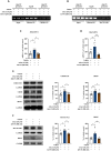
Fig. 1.. Inhibition of CREB (S133) by 666-15 reduces antidepressant-like effects of arketamine.
(A) Experimental design and timeline. CSDS was applied from days 1 to 10. On day 11, social interaction test (SIT) identified CSDS-susceptible mice. On day 12, mice received i.c.v. injections of 666-15 (CREB-CBP inhibitor) or vehicle, followed immediately by intraperitoneal (ip) injection of arketamine or vehicle. Behavioral assays: LMT, FST, and 1% SPT were conducted on subsequent days. (B to D) Results of the LMT, FST, and 1% SPT. Data are presented as mean ± SEM (n = 10 to 12). *P < 0.05, **P < 0.01, and ***P < 0.001 [one-way analysis of variance (ANOVA)]. (E) ChIP assay showing CREB (S133) binding to the Bdnf exon IV promoter in mPFC of mouse samples. (F) Bdnf mRNA levels in the mPFC. Data are presented as mean ± SEM (n = 6). *P < 0.05 and **P < 0.01 by one-way ANOVA. (G) The CREB (S133), CREB, and BDNF protein levels in the mPFC. Data are presented as mean ± SEM (n = 9). *P < 0.05 and **P < 0.01 by one-way ANOVA. GAPDH, glyceraldehyde phosphate dehydrogenase; NS, not significant.
Fig. 2.
Fig. 2.. Inhibition of MeCP2 S421 by KN-93 reduces antidepressant-like effects of arketamine.
(A) Experimental design and timeline. Mice underwent CSDS on days 1 to 10. On day 11, SIT identified CSDS-susceptible mice. On day 12, mice received i.c.v. injections of KN-93 (CaMKII inhibitor) or vehicle, immediately followed by intraperitoneal injection of arketamine or vehicle. Behavioral assays: LMT, FST, and 1% SPT were conducted on subsequent days. (B to D) Results of the LMT, FST, and 1% SPT. Data are presented as mean ± SEM (n = 9 or 10). *P < 0.05, **P < 0.01, and ***P < 0.001 (one-way ANOVA). (E) Binding affinity of MeCP2 (S421) to Bdnf IV promoters in the mPFC of mouse samples. (F) Bdnf mRNA levels in the mPFC. Data are presented as mean ± SEM (n = 6). *P < 0.05 and **P < 0.01 (one-way ANOVA). (G) The MeCP2 (S421) and BDNF protein levels in the mPFC. Data are presented as mean ± SEM (n = 9). *P < 0.05 and **P < 0.01 (one-way ANOVA).
Fig. 3.
Fig. 3.. Mutations of CREB at S133 and MeCP2 at S421 reduce the antidepressant-like effects of arketamine.
(A) Experimental design and timeline. On day 0, the virus was injected into the IL of the mPFC. CSDS was applied from days 11 to 20. On day 21, the SIT was performed to identify CSDS-susceptible mice. On day 22, arketamine or vehicle was bilaterally injected into the IL of mPFC, followed by behavioral tests, including the LMT, FST, and 1% SPT. (B) Illustration of the virus injection site in the mPFC (IL) and distribution of the virus in the brain. Scale bar, 100 μm. (C) Western blot analysis of BDNF protein levels in the mPFC of virus-injected mice (n = 6). *P < 0.05 and **P < 0.01 (one-way ANOVA). (D to F) Results of the LMT, FST, and 1% SPT. Data are presented as mean ± SEM (n = 16 to 18). **P < 0.01 and ***P < 0.001 (one-way ANOVA). (G) Binding affinity of CREB (S133) and MeCP2 (S421) to the Bdnf IV promoters in the mPFC of mouse samples. (H) Bdnf mRNA levels in the mPFC. Data are presented as mean ± SEM (n = 6). **P < 0.01 and ***P < 0.001 (one-way ANOVA). (I) The BDNF protein levels in the mPFC. Data are presented as mean ± SEM (n = 12). *P < 0.05, **P < 0.01, and ***P < 0.001 (one-way ANOVA).
Fig. 4.
Fig. 4.. 666-15 and KN-93 reduce BDNF transcription on arketamine-treated BV2 cells.
(A and B) Binding affinity of CREB (S133) and MeCP2 (S421) to Bdnf IV promoters on BV2 cells. (C and D) Bdnf mRNA levels on BV2 cells. Data are presented as mean ± SEM (n = 7 or 8). *P < 0.05, **P < 0.01, and ***P < 0.001 (one-way ANOVA). (E) The CREB (S133), CREB, and BDNF protein levels on BV2 cells. Data are presented as mean ± SEM (n = 8). *P < 0.05 and **P < 0.01 (one-way ANOVA). (F) The MeCP2 (S421) and BDNF protein levels on BV2 cells. Data are presented as mean ± SEM (n = 7). *P < 0.05 and **P < 0.01 (one-way ANOVA).
Fig. 5.
Fig. 5.. 666-15 and KN-93 reduce BDNF expression on arketamine-treated primary neurons.
(A) Coculture of primary microglia and primary neurons. (B) Bdnf mRNA levels on primary neurons. Data are presented as mean ± SEM (n = 6). ***P < 0.001 (one-way ANOVA). (C) The BDNF protein levels on primary neurons. Data are presented as mean ± SEM (n = 6). **P < 0.01 (one-way ANOVA). (D) Immunofluorescent staining for IBA1, NeuN, and BDNF on primary neurons and microglia. Scale bars, 25 μm.
Fig. 6.
Fig. 6.. Arketamine restores the reduced Bdnf mRNA levels in microglia of CSDS-susceptible mice.
(A) Schematic representation of the microglia isolation process from the mouse cerebral cortex. (B) Purity of isolated microglia confirmed using flow cytometry. SSC-A, Side Scatter Area; FSC-A, Forward Scatter Area; PE-A, Phycoerythrin Area. (C) Bdnf mRNA levels on microglia from the mouse cerebral cortex. Data are presented as mean ± SEM (n = 6). *P < 0.05 (one-way ANOVA). (D and E) Detection of Bdnf in mPFC microglia (Iba1+ cells) using Bdnf-RNAscope. Data are presented as mean ± SEM (n = 6). ***P < 0.001 (one-way ANOVA). Scale bar, 25 μm.
Fig. 7.
Fig. 7.. BDNF knockdown in mPFC microglia reduces the antidepressant-like effects of arketamine.
(A) Experimental timeline. On day 0, the virus was injected into the IL region of the mPFC. Tamoxifen was administered on days 3, 5, 7, 9, and 11. CSDS was applied from days 12 to 21. On day 22, the SIT identified CSDS-susceptible mice. On day 23, arketamine or vehicle was bilaterally injected into the mPFC (IL), followed by behavioral tests. (B) Diagram of the virus injection site in the mPFC (IL) and virus distribution. Scale bar, 25 μm. (C) Western blot analysis of BDNF protein expression in the mPFC (n = 6). ***P < 0.001 (Student’s t test). (D to F) Results of the LMT, FST, and 1% SPT. Data are presented as mean ± SEM (n = 12 or 13). *P < 0.05, **P < 0.01, and ***P < 0.001 (one-way ANOVA). (G) Schematic of whole-cell patch-clamp recording configuration of mPFC (IL) neurons from Cx3cr1-CreER mice receiving an intra-IL injection of the PLVX-CAG-DIO-shBDNF-mCherry or PLVX-CAG-DIO-mCherry. (H) Representative traces of sEPSCs recorded in mPFC (IL) neurons. Scale bars, 2 s, 10 pA. (I) Cumulative probability plots of sEPSC interevent intervals and average sEPSC frequency. *P < 0.05 and **P < 0.01 (one-way ANOVA). (J) Cumulative probability plots of sEPSC amplitudes and average sEPSC amplitudes. Data are presented as mean ± SEM (n = 20, 17, and 19 neurons from six mice). *P < 0.05 and **P < 0.01 (one-way ANOVA). (K) Voltage traces evoked by inward current injections (250, 275, and 300 pA) in mPFC (IL) neurons. Scale bars, 200 ms, 200 mV. (L) Plot of firing frequency versus depolarizing currents. Data are presented as mean ± SEM (n = 20, 17, and 18 neurons from six mice). *P < 0.05 and **P < 0.01 (two-way ANOVA). shRNA, short hairpin RNA.
Fig. 8.
Fig. 8.. Knockdown of TrkB in the mPFC reduces the antidepressant-like effects of arketamine.
(A) Experimental design and timeline. On day 0, the virus was injected into the IL region of the mPFC. CSDS was applied from days 11 to 20. On day 21, the SIT identified CSDS-susceptible mice. On day 22, arketamine or vehicle was bilaterally injected into the mPFC (IL). (B) Schematic representation of the IL injection site and virus distribution in the brain. Scale bar, 100 μm. PrL, Prelimbic Cortex; DP, Dorsal Prefrontal Cortex. (C) Western blot analysis of TrkB protein expression in the mPFC of virus-injected mice (n = 6). ***P < 0.001 (Student’s t test). (D to F) Results of the LMT, FST, and SPT. Data are presented as mean ± SEM (n = 9 or 10). *P < 0.05, **P < 0.01, and ***P < 0.001 (one-way ANOVA). (G) Schematic of whole-cell patch-clamp recording configuration of mPFC (IL) neurons from C57BL/6 mice receiving an intra-IL injection of the AAV-CaMKllα-shTrkB-mCherry or AAV-CaMKllα-mCherry. (H) Representative traces of sEPSCs in mPFC (IL) neurons. Scale bars, 2 s, 10 pA. (I) Cumulative probability plots of sEPSC interevent intervals and average sEPSC frequency. *P < 0.05 (one-way ANOVA). (J) Cumulative probability plots of sEPSC amplitude and average sEPSC amplitude. Data are presented as mean ± SEM (n = 19, 18, and 16 neurons from 6 mice). *P < 0.05 (one-way ANOVA). (K) Voltage traces evoked by inward current injections (250, 275, and 300 pA) in mPFC (IL) neurons. Scale bars, 200 ms, 200 mV. (L) Firing frequency as a function of depolarizing currents. Data are presented as mean ± SEM (n = 23, 20, and 20 neurons from six mice). *P < 0.05 (two-way ANOVA).
Fig. 9.
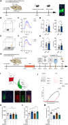
Fig. 9.. Disruption of the mPFC-NAc circuit diminishes the antidepressant-like effects of arketamine.
(A) Schematic representation of the experimental design. Scale bar, 100 μm. (B and E) Diagram of the fiber photometry apparatus used to measure Ca2+ signals in response to air puff and foot shock stimuli in mice treated with either vehicle or arketamine. (C and F) Averaged Ca2+ responses to air puff and foot shock stimuli in the mPFC (IL) of vehicle- or arketamine-treated mice. (D and G) Comparison of ΔF/F and the area under the curve (AUC) of ΔF/F for air puff and foot shock stimuli in the mPFC (IL) of vehicle- or arketamine-treated mice. Data are presented as mean ± SEM (trial = 24, mice = 6). *P < 0.05, **P < 0.01, and ***P < 0.001(Student’s t test). (H) Timeline of treatment and behavioral assessments. On day 0, a virus was injected into the mPFC (IL) and NAc (shell). CSDS was applied from days 14 to 24. On day 25, the SIT identified CSDS-susceptible mice. On day 26, arketamine or vehicle was bilaterally injected into the mPFC (IL). Behavioral tests, including the LMT, FST, and 1% SPT, were conducted thereafter. (I) Illustration of the IL injection site and virus distribution in the brain. Scale bar, 100 μm. eGFP, enhanced green fluorescent protein. (J) Inhibition of NAc (shell)–projecting mPFC (IL) neurons expressing hM4Di upon bath application of CNO (5 μM). Data are presented as mean ± SEM (n = 8 cells per group from three mice). ***P < 0.001 (two-way ANOVA). (K to M) Results of LMT, FST, and SPT. Data are presented as mean ± SEM (n = 12 or 14). *P < 0.05 and ***P < 0.001 (one-way ANOVA).

References

    1. Berman R. M., Cappiello A., Anand A., Oren D. A., Heninger G. R., Charney D. S., Krystal J. H., Antidepressant effects of ketamine in depressed patients. Biol. Psychiatry 47, 351–354 (2000). - PubMed
    1. Wilkinson S. T., Ballard E. D., Bloch M. H., Mathew S. J., Murrough J. W., Feder A., Sos P., Wang G., Zarate C. A. Jr., Sanacora G., The effect of a single dose of intravenous ketamine on suicidal ideation: A systematic review and individual participant data meta-analysis. Am. J. Psychiatry 175, 150–158 (2018). - PMC - PubMed
    1. Zarate C. A. Jr., Singh J. B., Carlson P. J., Brutsche N. E., Ameli R., Luckenbaugh D. A., Charney D. S., Manji H. K., A randomized trial of an N-methyl-D-aspartate antagonist in treatment-resistant major depression. Arch. Gen. Psychiatry 63, 856–864 (2006). - PubMed
    1. Zhang J. C., Yao W., Hashimoto K., Arketamine, a new rapid-acting antidepressant: A historical review and future directions. Neuropharmacology 218, 109219 (2022). - PubMed
    1. Hashimoto K., Molecular mechanisms of the rapid-acting and long-lasting antidepressant actions of (R)-ketamine. Biochem. Pharmacol. 177, 113935 (2020). - PubMed

MeSH terms

Substances