. 2025 Jul 9;16(1):506.
doi: 10.1038/s41419-025-07820-9.

M6A-METTL3-dependent nuclear PANC754/PSPC1/H3K4me1 repression complex regulate immune evasive LGALS7 signal to enhance immunotherapy against colorectal cancer

Affiliations

M6A-METTL3-dependent nuclear PANC754/PSPC1/H3K4me1 repression complex regulate immune evasive LGALS7 signal to enhance immunotherapy against colorectal cancer

Jianfeng Zhang et al. Cell Death Dis. .

Abstract

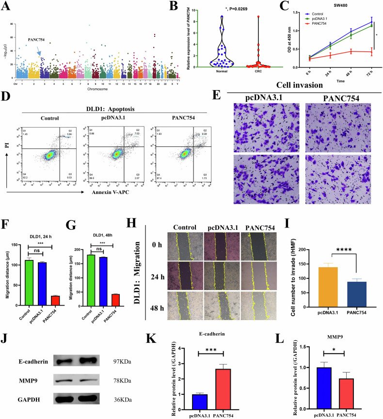
Non-coding RNAs (ncRNAs) have important regulatory functions similar to traditional oncogenes or tumor suppressor genes. Our previous research found a novel pan-cancer downexpressed ncRNA, PANC754. However, its function and underlying mechanism remain obscure in colorectal cancer (CRC). In this study, in vitro and in vivo experiments were performed to determine the function of PANC754. Loss and gain of function experiments, molecular docking experiments, and bioinformatic analysis were utilized to visualize its pathway. Co-culture system was leveraged to explore its effect on synergetic immune checkpoint blockage against CRC. Through a series of studies, we found that overexpressed PANC754 significantly inhibited cell viability, migration, and metastasis and induced notable apoptosis in CRC. The mechanical research found that PANC754 was the nuclear-located and its expression was regulated by m6A modification via METTL3 enzyme, which bound with its RBP PSPC1, then interacted with H3K4me1 to chromatin-accessible inhibit immune evasive molecule LGALS7 and led to suppress CRC progress. Furthermore, we confirmed that prominent upregulation of the immune checkpoint inhibitory (ICI) capability of anti-NKG2A, monalizumab when it was combined with PANC754 overexpression. Collectively, our study revealed that PANC754 is a tumor-suppressing ncRNA to form an ncRNA/RBP/histone repression complex with m6A-dependence, which can enhance the immune therapeutics effect of ICI, suggesting a promising therapeutic target. PANC754 is a novel pan-tumor suppressing non-coding RNA which is m6A-dependent and regulated by METTL3 modification enzyme. PANC754 is located at cellular nuclear and interacts with its RNA binding protein (RBP) PSPC1 and chromatin accessible histone H3K4me1, then can enhance immunotherapy capability of ICB anti-NKG2A against colorectal cancer through the immune evasive molecule LGALS7 signaling. Effect is that novel nuclear PANC754/PSPC1/H3K4me1 repression complex down-regulates the level "Don't eat me" signal LGALS7 to improve the immune efficiency of ICB and induce NK or CTL cell to release perforin and cytokine to kill tumor cells. (Created by Figdraw).


Conflict of interest statement

Competing interests: The authors declare no competing interests. Ethics approval and consent to participate: The studies involving human participants were ethically reviewed and approved by the Institutional Review Board of Affiliated Hospital of Nantong University (No. 2018-K008). The patients/participants provided their written informed consent to participate in this study. The animal experiments were ethically inspected and approved by the Laboratory Animal Ethical Committee of Nantong University (No. S20210301-020).

Figures

None
PANC754 is a novel pan-tumor suppressing non-coding RNA which is m6A-dependent and regulated by METTL3 modification enzyme. PANC754 is located at cellular nuclear and interacts with its RNA binding protein (RBP) PSPC1 and chromatin accessible histone H3K4me1, then can enhance immunotherapy capability of ICB anti-NKG2A against colorectal cancer through the immune evasive molecule LGALS7 signaling. Effect is that novel nuclear PANC754/PSPC1/H3K4me1 repression complex down-regulates the level “Don’t eat me” signal LGALS7 to improve the immune efficiency of ICB and induce NK or CTL cell to release perforin and cytokine to kill tumor cells. (Created by Figdraw).
Fig. 1
Fig. 1. PANC754 markedly inhibited the cell growth and metastasis of CRC cell lines.
A Manhattan map of PANC754 crosses 23 types of human cancers. Cancer samples were collected from the TCGA project (N = 10,490). Gene expression level was log2 transformed before the meta-analysis. Random effect models were applied for the aggregation. B The expression levels of PANC754 were validated in the tumor tissues and paratumorous tissues of 26 pairs of colorectal cancer patients by quantitative PCR. C The proliferative curve of SW480 cell line with overexpression of PANC754 by CCK-8 assay. Control, untransfected SW480; pcDNA3.1, empty pcDNA3.1 plasmid transfected; PANC754, overexpression plasmid of PANC754 transfected; *P < 0.05. D Cell apoptosis detection by flow cytometry. Each experiment was repeated at least three times. EG Determination of cell migration in DLD cell line by wound healing repair assay and its statistical histograms in 24 h F or 48 h. G ns, no significance; ***P < 0.001. H, I Cell invasion detection by transwell chamber assay and their statistical histograms in SW480 cell line. ****P < 0.0001. JL MMP9 and EMT marker E-cadherin were detected by WB and their statistical histograms in the SW480 cell line. Each experiment was repeated at least three times.
Fig. 2
Fig. 2. PANC754 significantly suppressed cell growth and metastasis in CDX model.
A The pipeline of CRC HCT116 cell line-derived xenograft (CDX; Created by Figdraw). B The presentation of the CDX tumor sizes in three different treatment groups (n = 5, respectively). WT, lentivirus uninfected SW480; Empty, empty lentivirus-infected; PANC754, overexpression PANC754 lentivirus-infected. C The CDX tumor volumes in three treated CDX groups. Empty vector, empty lentivirus vector infected; OE-PANC754, overexpression PANC754 lentivirus vector infected. **P < 0.01. D The representative image of H&E staining in three treatment CDX groups. E The representative image of IHC staining of the proliferation marker Ki67 in three different treatment groups. FH The protein levels of the metastasis biomarkers MMP9 and E-cadherin in CDX were detected by WB and their statistical histograms. ***P < 0.001.
Fig. 3
Fig. 3. PANC754 was regulated by m6A modification with methyltransferase METTL3.
A Prediction of m6A site of PANC754 in the SRAMP online website. High combined score implies high confidence. B Prediction of PANC754 secondary structure including m6A site in the SRAMP online website. C The schematic diagram of meRIP-PCR assay (Created with Microsoft PPT, and some icons in the schematic are sourced from bersinbio.com). D The m6A level of PANC754 was detected by meRIP-PCR. IgG served as a negative control. ***P < 0.001. E The m6A level of PANC754 in various CRC cell lines by meRIP-PCR. F The positive correlation between the m6A level and the mRNA level of PANC754. G With the knockdown of methyltransferase METTL3, the m6A level of PANC754 was markedly decline. H, I The mRNA level of PANC754 was significantly increase (H) or significantly decrease (I) after the overexpression or down-regulation of METTL3. NC, empty vector or scramble shRNA vector control; OE-METTL3, overexpression of METTL3; shMETTL3, down-regulation of METTL3; **P < 0.01; ****P < 0.0001; ns no significance; Each experiment was repeated at least three times.
Fig. 4
Fig. 4. PANC754 was the nuclear-located and bound with its RBP PSPC1.
A The subcellular location of PANC754 was detected by RNA FISH. U6 served as a nucleus location control; 18S served as a cytoplasm location biomarker. B The subcellular location of PANC754 by the nuclear-cytoplasmic fractionation experiment. GAPDH served as a cytoplasmic location biomarker. C The flowchart of RNA pulldown of PANC754 and subsequent analysis (Created with Microsoft PPT, and some icons in the schematic are sourced from bersinbio.com). D The agarose gel electrophoresis map verified that in vitro transcription approach to produce both sense and antisense RNA transcripts of PANC754. E The silver staining pattern of RNA pulldown between sense and antisense RNA transcripts of PANC754. F The Venn diagram of LS-MS/MS analysis between the sense and antisense RNA transcripts of PANC754. G Immunoblotting of the RNA pulldown samples further confirmed PSPC1 as the RBP for PANC754. H The mRNA level of PSPC1 in various CRC cell lines. I The mRNA level of PSPC1 when overexpression of PANC754 and/or knockdown of PSPC1. NC, HCT116 cells with untransfected plasmid; OE-PANC754, overexpression PANC754 plasmid transfected; shPSPC1, shPSPC1 plasmid transfected.
Fig. 5
Fig. 5. PANC754 binding with its RBP PSPC1 suppresses CRC progress via inhibiting immune evasive molecule LGALS7.
A The heat map of PANC754 overexpression in DLD1 cell line. NC, HCT116 cells with untransfected plasmid; 754, overexpression PANC754 plasmid transfected. B The volcano plot of PANC754 overexpression, where the arrow pointed LGALS7 significantly downregulated. C GSEA (gene set enrichment analysis) indicated that PANC754 overexpression led to the enrichment of the immune signals of leukocyte acitvation. D The mRNA levels of PANC754, PSPC1, and LGALS7 gene after PANC754 overexpression in DLD1 cells were detected by qRT-PCR. *P < 0.05; **P < 0.01; ***P < 0.001. Each experiment was repeated at least three times. E The mRNA levels of METTL3, PANC754, PSPC1, and LGALS7 gene after METTL3 overexpression in HCT16 cells were detected by qRT-PCR. F The mRNA levels of METTL3, PANC754, PSPC1, and LGALS7 gene after METTL3 knockdown in SW480 cells were detected by qRT-PCR. G The protein expression of PSPC1 and LGALS7 was determinedin SW480 cells by Western blotting. GAPDH served as the loading control; Empty vector, transfected with empty pcDNA3.1 plasmid group; OE-PANC754, transfected with pcDNA3.1-PANC754 overexpression plasmid group; shPANC754, transfected with shPANC754 kncokdown plasmid group.
Fig. 6
Fig. 6. PANC754/PSPC1/H3K4me1 repression complex regulates LGALS7 expression.
A The protein-protein interaction (PPI) diagram from RNA-Seq data suggests a strong correlation between PANC754’s downstream effects and the histone-encoding gene H3-4. B, C The histone H3K4me1 protein level after PANC754 overexpression in SW480 cells was detected WB and their statistic histograms. C PCNA was served a internal control. ***P < 0.001. Each experiment was repeated at least three times. D The interplay between PSPC1 and H3K4me1 by molecular docking experiment. E The direct interaction between PSPC1 and H3K4me1 was detected in Caco2 cells by Co-IP. F By using H3K4 inhibitor MTA, the mRNA level of LGALS7 gene decreasing slowly company with the increasing concentration of MTA in DLD1 cells. G A schematic diagram showed the model of m6A-dependent nuclear ncRNA PANC754 coupled with its binding protein PSPC1 and chromatin-accessible H3K4me1 protein to form ncRNA/RBP/histone repression complex to downregulate the immune invasive LGALS7 signaling, inhibiting colorectal cancer progress (Created with Microsoft Visio).
Fig. 7
Fig. 7. Upregulation of the immunotherapeutic ability of Monalizumab by PANC754 combination.
A The flowchart of co-culture system of PANC754 and the immune checkpoint blockage (ICB) of NKG2A gene, Monalizumab (Created by Figdraw). B The Perforin content in the culture supernatant of co-culture system was detected by ELISA. PF, Perforin; CRC, HCT116 cells; Control, transfected with empty plasmids; PBMC, peripheral blood mononuclear cells; OE-PANC754, transfected with PANC754 overexpression plasmid. C The apoptosis rate of HCT116 cells was detected by FCM. *P < 0.05; **P < 0.01; ***P < 0.001; ns, no significance. D The LDH concentration in the culture supernatant detected by velocity method in the biochemical analyzer. E The LDH concentration in the culture supernatant detected by ELISA. Gzms-B, granzyme B. F E-cadherin protein level in HCT116 cells was determined by immunocytochemical (ICC) staining. G The positive rate of MMP9 protein in HCT116 cells was determined by ICC staining. H The mRNA level of LGALS7 gene in HCT116 cells was determined by qRT-PCR. ****P < 0.0001. Each experiment was repeated at least three times.
Fig. 8
Fig. 8. cfPANC754 may serve as a biomarker for the diagnosis of colorectal cancer.
A The expression level of the cell-free RNA PANC754 in the serum (cfPANC754) from 75 CRC patients and 38 healthy controls was detected by QPCR. CRC, colorectal cancer; HC, healthy control. B The survival prediction ability of cfPANC754 by the total survival curve. High, high level of cfPANC754; low, low level of cfPANC754. C The graphical abstract of our research indicated that m6A/METTL3-dependent nuclear ncRNA PANC754 interacts with its binding protein PSPC1 and chromatin-accessible histone H3K4me1 to form ncRNA/RBP/histone repression complex near LGALS7’s promoter region can enhance immunotherapy capability of ICB anti-NKG2A against colorectal cancer through downregulation of the immune evasive LGALS7 signaling (Created by Figdraw).

References

    1. Yao RW, Wang Y, Chen LL. Cellular functions of long noncoding RNAs. Nat Cell Biol. 2019;21:542–51. - PubMed
    1. Slack FJ, Chinnaiyan AM. The role of non-coding RNAs in oncology. Cell. 2019;179:1033–55. - PMC - PubMed
    1. Decoding noncoding RNAs. Nat Methods. 2022;19:1147–8. - PubMed
    1. Palazzo AF, Lee ES. Non-coding RNA: what is functional and what is junk? Front Genet. 2015;6:2. - PMC - PubMed
    1. Ding W, Pu W, Jiang S, Ma Y, Liu Q, Wu W, et al. Evaluation of the antifibrotic potency by knocking down SPARC, CCR2 and SMAD3. EBioMedicine. 2018;38:238–47. - PMC - PubMed

MeSH terms