. 2025 Jul 9;16(1):508.
doi: 10.1038/s41419-025-07827-2.

Estrogen rescues muscle regeneration impaired by DUX4 in a humanized xenograft mouse model

Affiliations

Estrogen rescues muscle regeneration impaired by DUX4 in a humanized xenograft mouse model

Silvia Maiullari et al. Cell Death Dis. .

Abstract

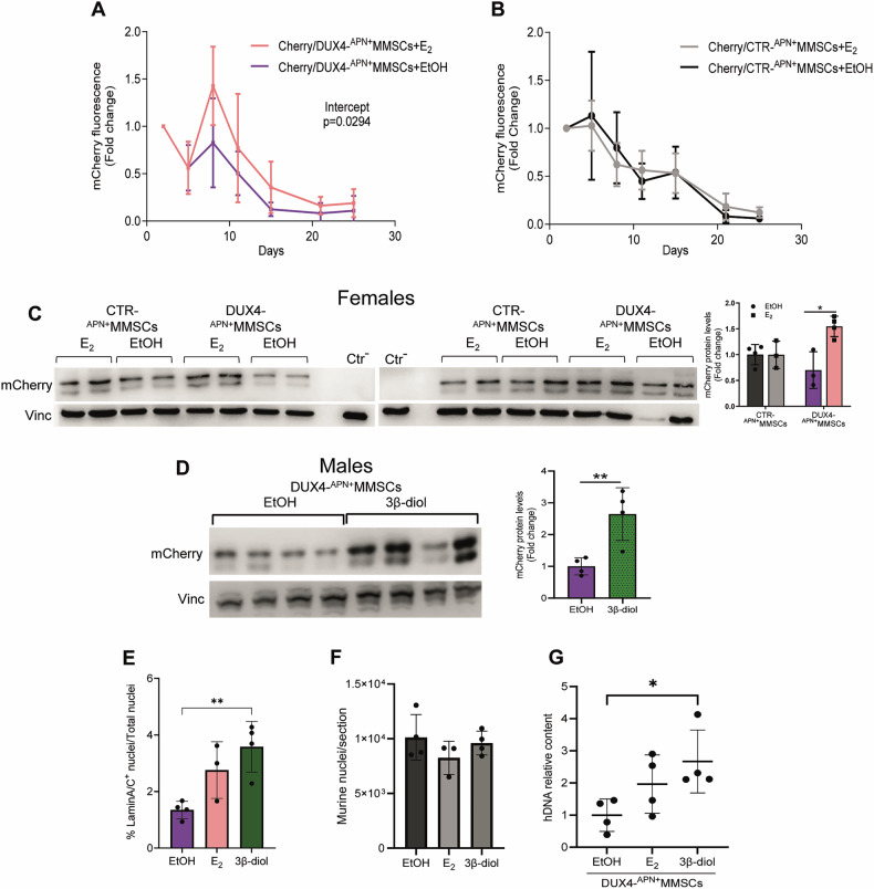
Facioscapulohumeral dystrophy (FSHD) is an autosomal dominant muscular dystrophy and one of the most frequent hereditary myopathies. The pathology shows a wide range of clinical signs, with modifying factors contributing to this variability, especially in patients with mild disease. Among these factors, the beneficial activity of estrogen hormones is controversial. We investigated the effect of 17β-estradiol (E2) and the 5α-dihydrotestosterone-derived 3β-androstenediol (3β-diol) on muscle regeneration. To recapitulate human hormone sensitivity, we developed a humanized heterokaryon FSHD mouse model by engrafting human immortalized myoblasts or human primary muscle mesenchymal stromal cells into surgically treated murine muscle. Inducible lentiviral expression of the pathogenic FSHD gene, DUX4, in human cells impaired the structural and functional recovery of murine muscle, providing a humanized mouse model of DUX4-mediated pathogenicity and proving that the biological effect of DUX4 spreads across the neighbouring murine nuclei. Both hormones counteracted DUX4 transcriptional activity and rescued structural and functional muscle performance impaired by DUX4 expression, while being inefficient in control grafts. The beneficial activity of estrogen in this heterokaryon model supports the hypothesis that these hormones contribute as a modifying factor in FSHD.


Conflict of interest statement

Competing interests: The authors declare no competing interests. Ethics approval and consent to participate: The collection of human biopsies was approved by the Ethical Committee of the Fondazione Policlinico Universitario Agostino Gemelli IRCCS, Università Cattolica del Sacro Cuore (Prot. ID 1524), and obtained after written informed consent from all participants. The study was performed in accordance with the Declaration of Helsinki. Animal studies obtained ethical approval from the Ministry of Health (Protocol N° 999/2017-PR) and were conducted in compliance with the institutional guidelines in accordance with Italian laws (DL N116, GU, suppl. 40, 18-2-1992).

Figures

Fig. 1
Fig. 1. DUX4 is active in human MMSCs.
Heat map (A) and cumulative graph (B) of alkaline phosphatase (ALP), platelet-derived growth factor receptor beta (PDGFRβ), neural/glial antigen 2 (NG2) (only in A), and platelet-derived growth factor receptor alpha (PDGFRα or CD140α) positive human MMSCs isolated from different healthy subject biopsies (CTR). Human fibroblasts (hFibr) and myoblasts (hMyob) served as controls. C DUX4 mRNA levels by RT-qPCR in APN+MMSCs from biopsies of 1 CTR and 3 FSHD1 patients. APN+MMSCs were collected in growth (GM) or differentiating medium (DM) at the indicated time points. All samples were normalized to hGAPDH and relative to the DUX4 value in GM FSHD1#1, set to 1. D Representative image of TUNEL+ DUX4-APN+MMSCs infected with mCherry and doxycycline-inducible DUX4 lentiviral vectors upon Dox treatment under growing conditions. The graph on the right reports the percentage of TUNEL+ cells/field in GM or the differentiation medium (DM) at the indicated time points. At least 9 fields from 2 biological replicates were counted. Scale bar is 5 µm. E Percentage of MHC+ cells/field in DUX4-APN+MMSCs grown in DM for 5 days and treated ±Dox (1 µg/ml) (nuclei in MHC+ cells/total number of nuclei). Five fields from 2 biological replicates were counted. Data is shown as mean ± SD, *P < 0.05, unpaired t-test. F Fusion index in DUX4-APN+MMSCs grown in DM for 7 days and treated ±Dox (percentage of nuclei in MHC+myotube, where a myotube is a MHC+ cell with at least 3 nuclei). Ten fields from two biological replicates were counted. Data is shown as mean ± SD, *P < 0.05, unpaired t-test. G Quantification of ERβ protein levels in CTR-APN+MMSCs and human myoblasts from healthy subjects. MCF7 cells were used as a positive control, and their value was arbitrarily set to 1. The relative blots are shown in Fig. S1G. TRIM43 (H) and ZSCAN4 (I) mRNA levels by RT-qPCR in APN+MMSCs overexpressing DUX4 and collected in GM or after 48 h in DM and treated as indicated. CTR is non-transfected APN+MMSCs. All samples were normalized to hGAPDH. n = 3 replicates/condition. Data is shown as mean ± SD., **P < 0.01, one-way ANOVA with Dunnett’s multiple comparisons test.
Fig. 2
Fig. 2. DUX4 impairs the contribution of human cells to murine muscle regeneration.
A Percentage of human genome content in male CTR-APN+MMSCs or CTR-ImMyobs engrafts and collected at the indicated time points. Human genome was calculated relative to an absolute standard curve, using FOXP1 gene primer pairs. Data is from one experiment, n = 4 mice/group. Data is shown as mean ± SD. *P < 0.05, **P < 0.01, multiple unpaired t-test. B Representative images of TA muscle grafts obtained by transplantation of 1 × 106 CTR-APN+MMSCs or CTR-ImMyobs. Mice were sacrificed 4 weeks following transplantation. Laminin stains myofibers (red), human Lamin A/C stains human nuclei (green), and DAPI counterstains nuclei (blue). On the left, higher magnification panels. Scale bar of the right panels is 250 µm. C Human Myogenin (hMyog) mRNA levels by RT-qPCR in CTR-APN+MMSCs or CTR-ImMyobs grafts and collected 28 days after surgery. All samples were normalized to hGAPDH and relative to the mean value of a group of CTR-ImMyobs arbitrarily set to 1. Data is from 1 experiment, n = 4 mice/group. Data is shown as mean ± SD. D Summary of treadmill test data performed with male mice undergoing surgery and transplanted with sole matrigel (Sham) or CTR-APN+MMSCs or CTR-ImMyobs, according to the scheme reported below the graph. The reduction was calculated relative to the mean of a 3-day basal-run test for each mouse. Data is from three independent experiments for Sham and one for CTR- APN+MMSCs and -ImMyobs. nsham = 10, nAPN+MMSCs = 4, nImMyobs = 4. Data is shown as mean ± SD, ****P < 0.0001, mixed effects model ANOVA (cell type factor p < 0.0001, matching p < 0.0001, no equal variability assumption). E Scheme of animal treatment. Summary of treadmill test data performed with male animals undergoing surgery and transplanted with sole matrigel (Sham) or transplanted with CTR-APN+MMSCs or DUX4-APN+MMSCs (F) or CTR-ImMyobs or DUX4-ImMyobs (G). The reduction was calculated relative to the mean of a 3-day basal-run test for each mouse. Data is from three independent experiments for MMSCs nsham = 8, nCTR-MMSCs = 8, nDUX4-MMSCs = 6, and one experiment for ImMyobs. nCTR-ImMyobs = 4, nDUX4-ImMyobs = 4. Data is shown as mean ± SD. **P < 0.01, ***P < 0.001, mixed effects model ANOVA (F, cell type factor p = 0.0004, matching p < 0.0001; G, cell type factor p = 0.009, matching p < 0.0001, no equal variability assumption).
Fig. 3
Fig. 3. Estrogen antagonizes DUX4 transcriptional activity.
A Scheme of short-term animal treatment. Vehicle (EtOH), E2 (100 µg/kg body weight), 3β-diol (1 mg/kg bw), ICI (200 mg/kg bd). Human TRIM43 (B) and LEUTX (C) mRNA levels by RT-qPCR in male CTR- or DUX4- APN+MMSCs grafts treated as indicated. All samples were normalized to hAct and relative to EtOH-treated DUX4 transplanted animals, whose mean was arbitrarily set to 1. n = 3 mice/group. Data is shown as mean ± SD. Data is from 1 experiment, *P < 0.05, **P < 0.01, ***P < 0.001. One-way ANOVA with Dunnett’s multiple comparisons test. D Human DNA quantification by q-PCR in the TRIzol DNA fractions of the same samples as in (B, C). Human genome was calculated relative to an absolute standard curve, using FOXP1 gene primer pairs. n = 3 mice/group. E Human GAPDH mRNA levels by RT-qPCR in TA muscles of the same samples as in (A, B). Samples were normalized to murine GAPDH. Murine Wfdc3 mRNA levels by RT-qPCR in TA muscle of male DUX4- APN+MMSCs (F) or DUX4-ImMyobs grafts (G), treated as indicated. All samples were normalized to murine GAPDH. The Wfdc3 levels were relative to those of the contralateral leg of each mouse (F) or to EtOH-treated mice arbitrarily set to 1 (G). Data are from muscles as in (B, C), n = 3 mice/group (F). Data is from one experiment, n = 4 mice/group (G). Data is shown as mean ± SD. *P < 0.05, One-way ANOVA with Dunnett’s multiple comparisons test.
Fig. 4
Fig. 4. Estrogen antagonizes DUX4-mediated toxicity.
Quantification of mCherry fluorescence signal by TA muscle of female mice transplanted with DUX4-APN+MMSCs (A) or CTR-APN+MMSCs (B) and treated with E2 or EtOH according to the scheme in Fig. 2E. Photon emission was measured as photons per second (p/s). Data is from one experiment, n = 6 mice/group. Data is shown as mean ± SD. *P < 0.05, Linear regression: Intercept values, F = 4891 DFn = 1 DFd = 88. WB analysis of mCherry protein in DUX4- or CTR-APN+MMSCs grafts of female (C) animals as in A, B, or male animals (D) treated as indicated. Animals were sacrificed five (C) or 4 weeks (D) after transplantation. The graphs on the right report the relative quantification of WB. All samples were normalized to murine Vinculin. D n = 4, data is shown as mean ± SD. *P < 0.05, **P < 0.01. Unpaired t-test. Percentage of human nuclei (E) and total murine nuclei (F) in sequential sections of male DUX4-APN+MMSCs grafts, treated as indicated. Each data point is the mean of four different sections/murine TA encompassing the engraftment site (according to the scheme in Fig. S4D). Animals were sacrificed 4 weeks following transplantation. Data is a pool of two independent experiments, n ≧ 3 mice/group. Data is shown as mean ± SD, **P < 0.01, One-way ANOVA with Dunnett’s multiple comparisons test. G Human DNA quantification by qPCR in TRIzol DNA-fractions of male DUX4-APN+MMSCs grafts, treated as indicated, and sacrificed after 4 weeks. All samples were analysed using human TITIN gene and normalized to murine PTGER. Data is from one experiment, n = 4. Data is shown as mean ± SD, *P < 0.05, One-way ANOVA with Dunnett’s multiple comparisons test.
Fig. 5
Fig. 5. Estrogen rescues DUX4-impaired muscle running functionality and regeneration.
Summary of treadmill test data performed with male mice treated as indicated. The reduction was calculated relative to the mean of a 3-day basal-run test for each mouse. Data is a pool of two (A) or one experiment (B). DUX4-APN+MMSCs: nEtOH = 6, nE2 = 10, n3β-diol = 7. DUX4-ImMyobs: nEtOH = 4, nE2 = 4, n3β-diol = 6. Data is shown as mean ± SD. ****P < 0.0001, mixed effects model ANOVA (A, hormone treatment factor p < 0.0001, matching p < 0.0001; B, hormone treatment factor p < 0.0001, matching p < 0.0001, no equal variability assumption). C Treadmill data of healthy mice treated as indicated. Data is a pool of two experiments, nEtOH = 21, nE2 = 5, n3β-diol = 4. CSA distribution in the 10–8000 µm2 range of male mice transplanted with DUX4-APN+MMSCs (DF) or DUX4-ImMyobs (EG) and treated as indicated. Each bar is the mean of 4 mice. Four sections/mouse were evaluated using the MyoEngraftmentProfiler” pipeline (Fig. S6, see STAR Methods section). Data is a pool of two experiments. P < 0.05 Kolmogorov-Smirnov test. H Representative images of male DUX4-APN+MMSCs graft sections, treated as indicated. Animals were sacrificed 4 weeks following transplantation. Laminin stains myofiber (red) and DAPI counterstains nuclei (blue). Scale bar is 50 µm. Quantification of CNF (centrally nucleated myofibers in transversal sections, containing at least one central nucleus in TA muscle of male DUX4-APN+MMSCs (I) or female CTR- or DUX4-APN+MMSCs (J) grafts. Animals were sacrificed five (I) or 4 weeks (J) after transplantation. Data in I is a pool of three independent experiments. Data in J is from one experiment. n = 4 mice/group, 3 sections/mouse (I). n = 3 mice, 2 sections/mouse (J), Data is shown as mean ± SD. *P < 0.05, **P < 0.01, one-way ANOVA with Dunnett’s multiple comparisons test.
Fig. 6
Fig. 6. Estrogen rescues DUX4-induced muscle fibrosis.
A Representative images of muscle fibrosis in transversal sections of CTR- or DUX4-APN+MMSCs or matrigel (Sham) grafts and treated as indicated. Mice were sacrificed 4 weeks (APN+MMSCs) or 35 days (Sham) following transplantation. Images were obtained by picrosirius red staining. Quantification of muscle fibrosis in transversal sections of male (B, C) or female (D) grafts transplanted with indicated cells and treated as indicated. Data is expressed as a percentage of red picrosirius+ pixels to the total (red+yellow) pixel number. Data is from one CTR-APN+MMSCs or a pool of two DUX4-APN+MMSCs independent experiments (B, C) or one experimental group in D. Animals in B, C were sacrificed 4 weeks after transplantation, those in D 5 weeks after transplantation. nCTR-APN+MMSCs = 3 mice/group, 2 sections/mouse, nDUX4-APN+MMSCs≥3mice/group, 2 sections/mouse. nDUX4-ImMyobs = 4 mice/group, three sections/mouse. Data is shown as mean ± SD. *P < 0.05, ***P < 0.001. one-way ANOVA with Dunnett’s multiple comparisons test.

References

    1. Deenen JC, Arnts H, van der Maarel SM, Padberg GW, Verschuuren JJ, Bakker E, et al. Population-based incidence and prevalence of facioscapulohumeral dystrophy. Neurology. 2014;83:1956–9. - PMC - PubMed
    1. Hamel J, Tawil R. Facioscapulohumeral muscular dystrophy: update on pathogenesis and future treatments. Neurotherapeutics. 2018;15:863–71. - PMC - PubMed
    1. Daxinger L, Tapscott SJ, van der Maarel SM. Genetic and epigenetic contributors to FSHD. Curr Opin Genet Dev. 2015;33:56–61. - PMC - PubMed
    1. Lemmers RJ, van der Vliet PJ, Klooster R, Sacconi S, Camano P, Dauwerse JG, et al. A unifying genetic model for facioscapulohumeral muscular dystrophy. Science. 2010;329:1650–3. - PMC - PubMed
    1. Gabriëls J, Beckers MC, Ding H, De Vriese A, Plaisance S, van der Maarel SM, et al. Nucleotide sequence of the partially deleted D4Z4 locus in a patient with FSHD identifies a putative gene within each 3.3 kb element. Gene. 1999;236:25–32. - PubMed

MeSH terms

LinkOut - more resources