. 2025 Jul 9;16(1):6318.
doi: 10.1038/s41467-025-61401-0.

Mineralocorticoid receptor activation contributes to intestinal fibrosis through neutrophil gelatinase-associated lipocalin in preclinical models

Affiliations

Mineralocorticoid receptor activation contributes to intestinal fibrosis through neutrophil gelatinase-associated lipocalin in preclinical models

Asma Amamou et al. Nat Commun. .

Abstract

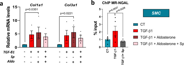
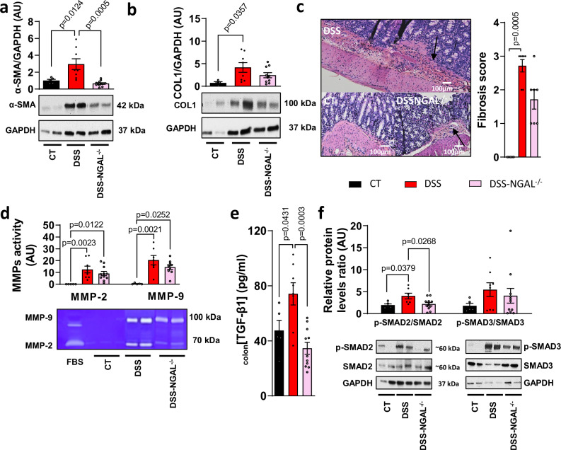
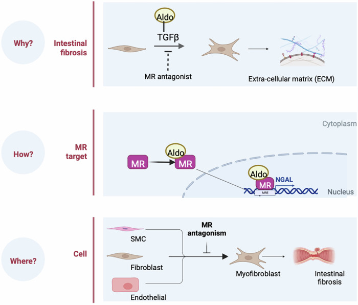
Intestinal fibrosis is a common complication in inflammatory bowel diseases with no specific therapy. Because mineralocorticoid receptor antagonism prevented inflammation and fibrosis in extra-intestinal organs, we aimed to evaluate mineralocorticoid receptor antagonism in intestinal fibrosis. Here we show that pharmacological or smooth cell specific deletion mineralocorticoid receptor antagonism prevented colon fibrosis development in male mice. In vitro, spironolactone prevented fibroblast proliferation and endothelial-to-mesenchymal transition. Neutrophil gelatinase-associated lipocalin silencing suppressed aldosterone-induced fibrosis markers and blunted colon fibrosis in mice. Chromatin immunoprecipitation showed mineralocorticoid receptor antagonist inhibits mineralocorticoid receptor binding on the neutrophil gelatinase-associated lipocalin promoter in activated smooth muscle cells. In conclusion, mineralocorticoid receptor antagonism or smooth muscle mineralocorticoid receptor deletion reduced colon fibrosis through the modulation of the neutrophil gelatinase-associated lipocalin pathway. Mineralocorticoid receptor may represent a novel therapeutic target in intestinal fibrosis and may allow the re-positioning in the field of inflammatory bowel diseases of drugs already marketed.


Conflict of interest statement

Competing interests: The authors declare no competing interests.

Figures

Fig. 1
Fig. 1. Dextran sulfate sodium (DSS)-induced chronic colitis and fibrosis upregulates plasma aldosterone and mineralocorticoid receptor (MR) activation.
Male C57BL/6 J mice underwent 3 cycles of 2% DSS in their drinking water for 7 days, followed by 14 days of regular water (DSS; n = 11) whereas control mice received normal water (CT; n = 10). a Experimental design. b Plasma aldosterone level (n = 10 for CT, n = 11 for DSS; Two-sided Mann-Whitney test). c, d Representative western blot and relative protein expression of colon MR (c, n = 10 for CT, n = 11 for DSS; Two-sided Mann-Whitney test) and SGK1 (d, n = 9 for CT, n = 12 for DSS; Two-sided unpaired t-test with Welch’s correction). GAPDH is used as an internal control. Data are presented as mean values +/− SEM. Created in BioRender.
Fig. 2
Fig. 2. Mineralocorticoid receptor (MR) antagonism by spironolactone decreases intestinal fibrosis in mice with chronic colitis.
a–f Pharmacological inhibition of MR by spironolactone in male mice with dextran sulfate sodium (DSS)-induced chronic colitis. Male C57BL/6 J mice underwent 3 cycles of 2% DSS in their drinking water for 7 days, followed by 14 days of regular water (DSS, n = 11) for both first cycles and 7 days for the third cycle whereas control mice received normal water (CT, n = 10). Mice were subjected to either a standard diet or diet supplemented with spironolactone (30 mg.kg−1; DSS+Sp, n = 12) throughout the experiment. (a) Colonic relative mRNA levels encoding for Tgfb1, Smad2, Smad3, Mmp3, Mmp9, Timp1 and Tnf (n = 9 for CT, n = 11 for DSS, n = 12 for DSS+Sp; ordinary one way ANOVA with Tukey’s multiple comparisons test). b Representative western blot and relative protein expression of colon COL1 (n = 10 for CT, n = 11 for DSS, n = 12 for DSS+Sp; ordinary one way ANOVA with Tukey’s multiple comparisons test). c Sirius red stained colon sections, fibrosis score and collagen area fraction (n = 9 for CT, n = 11 for DSS, n = 12 for DSS+Sp; Kruskal-Wallis test with Dunn’s multiple comparisons test). d Representative gelatin zymography and quantification of colon MMP-9 (n = 10 for CT, n = 11 for DSS, n = 12 for DSS+Sp; Kruskal-Wallis test with Dunn’s multiple comparisons test) and −2 (n = 10 for CT, n = 11 for DSS, n = 11 for DSS+Sp; ordinary one way ANOVA with Tukey’s multiple comparisons test) activities. e Colonic TGF-β1 level (n = 10 for CT, n = 11 for DSS, n = 12 for DSS+Sp; ordinary one way ANOVA with Tukey’s multiple comparisons test). (f) Representative western blot and relative protein expression of phospho-SMAD 2/3 (p-SMAD2, p-SMAD3) and SMAD 2/3 (n = 6 for CT, n = 8 for DSS, n = 8 for DSS+Sp; ordinary one way ANOVA with Tukey’s multiple comparisons test for p-Smad2/Smad2, Kruskal-Wallis test with Dunn’s multiple comparisons test for p-Smad3/Smad3). Data are presented as mean values +/− SEM.
Fig. 3
Fig. 3. Genetic smooth muscle deletion of mineralocorticoid receptor (MR) decreases intestinal fibrosis in mice with chronic colitis.
a–f Smooth muscle cell-specific inhibition of MR in female mice with dextran sulfate sodium (DSS)-induced chronic colitis. Female C57BL/6 J mice underwent 3 cycles of 2% DSS in their drinking water for 7 days, followed by 14 days of regular water for the both first cycles and 7 days for the third cycle (DSS-WT, n = 8; DSS-sm22MR−/−(KO), n = 10). a Colonic relative mRNA levels encoding for Tgfb1, Mmp3, Mmp9, Timp1, Vim, Fn1 and Tnf (n = 8 per group; Two-sided unpaired t-test with Welch’s correction for Mmp3, Mmp9, Timp1, Fn1 and Tnf; Two-sided unpaired t-test without Welch’s correction for Tgfb1 and Vim). b Representative western blot and relative protein expression of colon COL1 (n = 8 for DSS-WT, n = 10 for DSS-sm22MR−/−; Two-sided unpaired t-test with Welch’s correction). c Sirius red colon sections, fibrosis score and collagen area fraction (n = 7 for DSS-WT, n = 9 for DSS-sm22MR−/−; Two-sided Mann-Whitney test for fibrosis score; Two-sided unpaired t-test without Welch’s correction for collagen area fraction). d Representative gelatin zymography and quantification of colon MMP-9 and MMP-2 activity (n = 7 for DSS-WT, n = 10 for DSS-sm22MR−/−; Two-sided Mann-Whitney test). e Colonic TGF-β1 level (n = 8 for DSS-WT, n = 10 for DSS-sm22MR−/−; Two-sided unpaired t-test without Welch’s correction). f Representative western blot and relative protein expression of phospho-SMAD 2/3 (p-SMAD2, p-SMAD3) and SMAD 2/3 (n = 8 for DSS-WT, n = 10 for DSS-sm22MR−/−; Two-sided Mann-Whitney test). Data are presented as mean values +/− SEM.
Fig. 4
Fig. 4. Mineralocorticoid receptor (MR) activation exacerbates extracellular matrix fibroblast synthesis and promotes endothelial mesenchymal transition.
CCD-18Co (a–e) and HIMEC (f–j) cells were incubated with aldosterone (100 nM), spironolactone (10 µM) in response to TGF-β (10 ng/mL) for 24 h (n = 6 from independent experiments). (a) Relative mRNA levels of MR (Nr3c2) from CCD-18Co cell lysates (n = 6 per group; Two-sided Mann-Whitney test). b Relative mRNA levels of Acta2, Ccn2, and Col1a1 from CCD-18Co cell lysates (n = 6 per group; ordinary one-way ANOVA with Tukey’s multiple comparisons test). c Relative protein expression of α-SMA from CCD-18Co cell lysates (n = 6 for CT, n = 6 for TGF-β1, n = 5 for Aldosterone, n = 6 for TGF-β1 + Aldosterone, n = 6 for TGF−β1 + Aldosterone + Sp; ordinary one-way ANOVA with Tukey’s multiple comparisons test). d, e Representative gelatine zymography and quantification of MMP-2 activity from CCD-18Co cell lysates (n = 6 per group; ordinary one-way ANOVA with Tukey’s multiple comparisons test). (f) Relative mRNA levels of MR (Nr3c2) from HIMEC cell lysates (n = 5 per group; Two-sided Mann-Whitney test). g Relative mRNA levels of Acta2, Ccn2, and Col1a1 from HIMEC cell lysates (n = 6 per group; ordinary one-way ANOVA with Tukey’s multiple comparisons test). h Representative western blot and relative protein expression of α-SMA from HIMEC cell lysates (n = 5 per group; ordinary one-way ANOVA with Tukey’s multiple comparisons test). i, j Representative gelatin zymography and quantification of MMP-2 activity from HIMEC cell lysates (n = 5 per group; ordinary one-way ANOVA with Tukey’s multiple comparisons test). Data are presented as mean values +/− SEM.
Fig. 5
Fig. 5. Neutrophil gelatinase-associated lipocalin (NGAL) level is increased in colon of mice with chronic colitis and in aldosterone-induced colon fibroblasts.
a, b Male C57BL/6 J mice underwent 3 cycles of 2% dextran sulfate sodium (DSS) in their drinking water for 7 days, followed by 14 days of regular water (DSS, n = 11) for both first cycles and 7 days for the third cycle. whereas control mice received normal water (CT, n = 10). Mice were subjected to either a standard diet or diet supplemented with spironolactone (30 mg.kg-1; DSS+Sp, n = 12) throughout the experiment. a Relative mRNA and b protein expression of NGAL from colon of CT, DSS and DSS+Sp mice (n = 10 for CT, n = 11 for DSS, n = 12 for DSS+Sp; Kruskal-Wallis test with Dunn’s multiple comparisons test for mRNA expression; ordinary one way ANOVA with Tukey’s multiple comparisons test for protein expression). c–d Female C57BL/6 J mice underwent 3 cycles of 2% DSS in their drinking water for 7 days, followed by 14 days of regular water (DSS-WT, n = 8; DSS-sm22MR−/−, n = 10) for both first cycles and 7 days for the third cycle. c Relative mRNA and d protein expression of NGAL from colon of CT-WT, DSS-WT, DSS-sm22MR−/−mice (n = 8 for DSS = WT, n = 10 for DSS-sm22MR−/−; Two-sided unpaired t-test with Welch’s correction for mRNA epression; Two-sided Mann-Whitney test for protein expression). e Colon immunostaining of NR3C2, NGAL and SGK1 (n = 5 for CT, n = 10 for DSS, n = 12 for DSS+Sp; Kruskal-Wallis test with Dunn’s multiple comparisons test). f CCD-18Co cells were incubated with aldosterone (100 nM), spironolactone (10 µM) in response to TGF-β (10 ng/mL) for 24 h (n = 5 from independent experiments). Relative protein expression of NGAL from CCD-18Co cells lysates (n = 5 per group; ordinary one way ANOVA with Tukey’s multiple comparisons test). Data are presented as mean values +/− SEM.
Fig. 6
Fig. 6. Spironolactone inhibits mineralocorticoid receptor (MR) binding on the neutrophil gelatinase-associated lipocalin (NGAL) in TGF-β1-activated smooth muscle cells (SMC).
a, b Murine SMC were incubated with aldosterone (100 nM), spironolactone (10 µM) in response to TGF-β (10 ng/mL) for 24 h (n = 4 from independent experiments). a Relative mRNA expression of Cola1 and Col3a1 from SMC cells lysates (n = 4 per group; Kruskal-Wallis test with Dunn’s multiple comparisons test). b Chromatin immunoprecipitation (ChIP) followed by qPCR analysis (ChIP-qPCR) performed with an anti-MR antibody in SMC upon of TGF-β1 and spironolactone stimulation (n = 5 per group; Friedman test with Dunn’s multiple comparisons test). Data are presented as mean values +/− SEM.
Fig. 7
Fig. 7. Neutrophil gelatinase-associated lipocalin (NGAL) genetic deletion decreased intestinal fibrosis and TGF-β1 pathways in mice with chronic colitis.
Female C57BL/6 NGAL−/− (n = 13) and DSS (n = 8) underwent 3 cycles of 2% DSS in their drinking water for 7 days, followed by 14 days of regular for both first cycles and 7 days for the third cycle. Control mice received normal drinking water (CT; n = 10). a, b Representative western blot and relative protein level of α-SMA (a, n = 5 for CT, n = 8 for DSS, n = 11 for DSS-NGAL−/−; ordinary one way ANOVA with Tukey’s multiple comparisons test) and COL1 (b, n = 4 for CT, n = 8 for DSS, n = 10 for DSS-NGAL−/−; Kruskal-Wallis test with Dunn’s multiple comparisons test) colon. c Hematoxylin-eosin-safran stained tissues in CT, DSS, DSS-NGAL−/− groups and fibrosis score (n = 5 for CT, n = 7 for DSS, n = 7 for DSS-NGAL−/−; Kruskal-Wallis test with Dunn’s multiple comparisons test). d Representative gelatine zymography and quantification of colon MMP-9 (n = 5 for CT, n = 7 for DSS, n = 9 for DSS-NGAL−/−; Kruskal-Wallis test with Dunn’s multiple comparisons test) and MMP-2 (n = 5 for CT, n = 8 for DSS, n = 11 for DSS-NGAL−/−; Kruskal-Wallis test with Dunn’s multiple comparisons test) activity. e Colonic TGF-β1 level (n = 5 for CT, n = 8 for DSS, n = 11 for DSS-NGAL−/−; ordinary one-way ANOVA with Tukey’s multiple comparisons test). f Representative western blot and relative protein expression of phospho-SMAD 2/3 (p-SMAD2/SMAD2: n = 5 for CT, n = 7 for DSS, n = 11 for DSS-NGAL−/−, ordinary one way ANOVA with Tukey’s multiple comparisons test; p-SMAD3/SMAD3: n = 5 for CT, n = 8 for DSS, n = 11 for DSS-NGAL−/−; Kruskal-Wallis test with Dunn’s multiple comparisons test). Data are presented as mean values +/− SEM.
Fig. 8
Fig. 8. Neutrophil gelatinase-associated lipocalin (NGAL) mediates pro-fibrotic effect of mineralocorticoid receptor (MR) activation.
a, b CCD-18Co cells silenced for NGAL with siNGAL were incubated with aldosterone (Aldo, 100 nM) with or without TGF-β (10 ng/mL) for 24 h (n = 4 from independent experiments). Relative protein levels of α-SMA (a), COL1 (b) and representative image of western blot (c) from CCD-18Co cells lysates (n = 4 per group; Two-sided Mann-Whitney test). d–f CCD-18Co cells were incubated with increasing concentration of recombinant hNGAL (5 ng/ml and 50 ng/ml; n = 5 from independent experiments). d Relative mRNA level of Acta2, Ccn2 and Col1a1 from CCD-18Co cells lysates (n = 5 per group; ordinary one way ANOVA with Tukey’s multiple comparisons test). e Representative and relative protein expression of α-SMA from CCD-18Co cells lysates (n = 5 per group; ordinary one way ANOVA with Tukey’s multiple comparisons test). f Representative gelatine zymography and quantification of MMP-2 activity from CCD-18Co cell lysates (n = 5 per group; ordinary one way ANOVA with Tukey’s multiple comparisons test). Data are presented as mean values +/− SEM.
Fig. 9
Fig. 9. Effect of genetic deletion of neutrophil gelatinase-associated lipocalin (NGAL) on colon mineralocorticoid receptor (MR) and SGK1 expression in female mice with dextran sulfate sodium (DSS)-induced colitis.
Female C57BL/6 DSS-NGAL−/− (n = 13) and DSS (n = 8) underwent 3 cycles of 2% DSS in their drinking water for 7 days, followed by 14 days of regular water for both first cycles and 7 days for the third cycle. Control mice received normal water (CT; n = 10). a, b Representative western and relative protein level of MR (a) and SGK1 (b) colon (n = 5 for CT, n = 8 for DSS, n = 11 for DSS-NGAL−/−; Kruskal-Wallis test with Dunn’s multiple comparisons test). Data are presented as mean values +/− SEM.
Fig. 10
Fig. 10. Mineralocorticoid receptor (MR) activation promotes experimental intestinal fibrosis through neutrophil gelatinase-associated lipocalin (NGAL) pathway.
Aldosterone (Aldo), MR ligand promotes fibroblast activation while MR antagonism by spironolactone inhibited it. NGAL is a downstream MR target in intestinal fibrosis. MR antagonism reduced extracellular matrix production in smooth muscle cells (SMC), intestinal fibroblasts, and intestinal endothelial cells. Created in BioRender.

References

    1. D’Alessio, S. et al. Revisiting fibrosis in inflammatory bowel disease: the gut thickens. Nat. Rev. Gastroenterol. Hepatol.19, 169–184 (2022). - PubMed
    1. Rieder, F., Fiocchi, C. & Rogler, G. Mechanisms, management, and treatment of fibrosis in patients with inflammatory bowel diseases. Gastroenterology152, 340–350.e346 (2017). - PMC - PubMed
    1. D’Haens, G. et al. Challenges in the pathophysiology, diagnosis, and management of intestinal fibrosis in inflammatory bowel disease. Gastroenterology162, 26–31 (2022). - PMC - PubMed
    1. Vuyyuru, S. K. et al. Real world analysis on the efficacy and safety of anti-tumor necrosis factor therapy in patients with stricturing Crohn’s disease. Sci. Rep.11, 11704 (2021). - PMC - PubMed
    1. Moran, G. W. et al. Phenotypic features of crohn’s disease associated with failure of medical treatment. Clin. Gastroenterol. Hepatol.12, 434–442.e431 (2014). - PubMed

MeSH terms

LinkOut - more resources