. 2025 Nov;21(11):1762-1772.
doi: 10.1038/s41589-025-01974-5. Epub 2025 Jul 9.

Serine ADPr on histones and PARP1 is a cellular target of ester-linked ubiquitylation

Affiliations

Serine ADPr on histones and PARP1 is a cellular target of ester-linked ubiquitylation

Andreas Kolvenbach et al. Nat Chem Biol. 2025 Nov.

Erratum in

Abstract

ADP-ribosylation and ubiquitylation regulate various cellular processes, with the complexity of their interplay becoming increasingly clear, as illustrated by ADP-ribosylation-dependent ubiquitylation mediated by Legionella. Biochemical studies have reported ester-linked ubiquitylation of ADP-ribose by DELTEX ubiquitin ligases, yet the modification sites on cellular targets remain unknown. Here, our search for interactors of RNF114 revealed DNA-damage-induced serine mono-ADP-ribosylation as a cellular target for ester-linked ubiquitylation. By developing proteomics strategies tailored to the chemical features of this composite modification, combined with an enrichment method using the zfDi19 and ubiquitin interaction motif domain (ZUD) of RNF114 and specific chemical elution, we identified ADP-ribosyl-linked serine ubiquitylation sites in cells, including on histones and poly(ADP-ribose) polymerase 1. Engineering ZUD into a modular reagent enabled the detection of this dual modification by immunoblotting. We establish ADP-ribosyl-ubiquitylation as an endogenous serine post-translational modification and propose that our multifaceted, tailored methodology will uncover its widespread occurrence, along with other conjugation chemistries, across diverse signaling pathways.


Conflict of interest statement

Competing interests: I.M. declares that Max Planck Innovation, which is responsible for technology transfer from Max Planck Institutes, has licensed the antibody AbD43647 to Bio-Rad Laboratories, which markets it for research purposes. I.M., A.K. and M.D.P. are named as inventors on a pending patent application related to the work described in this manuscript. The remaining authors declare no competing interests. Inclusion & ethics statement: All authors fully fulfilled the criteria for authorship required by Nature Portfolio, as their participation was essential for the design and implementation of the study. Roles and responsibilities were agreed among authors ahead of the research. This research does not result in stigmatization, discrimination, incrimination or personal risk to researchers.

Figures

Fig. 1
Fig. 1. Identification of RNF114 interactors dependent on its zfDi19 domain and DNA damage.
a, Schematic representation of RNF114 WT domain structure and C176A mutant. RNF114 contains a RING finger and Zn1 domain required for catalytic activity. Zn2 and Zn3 form the zfDi19 domain required for mono-ADPr binding. A single substitution in the zfDi19 domain (C176A) abolishes mono-ADPr binding. The UIM binds ubiquitin. b, Experimental setup of the GFP pulldown. RNF114-KO U2OS cells complemented with inducible GFP–RNF114 WT or GFP–RNF114-C716A were left untreated or treated with 1 mM H2O2 for 1 h. Each condition consisted of four biological replicates. The pulldown was performed under nondenaturing conditions to identify interactors dependent on an intact zfDi19 domain and DNA damage, followed by MS analysis. c, Volcano plot showing the log2 fold change of the interactors identified in RNF114 WT-overexpressing cells comparing the DNA-damage-treated and untreated conditions. d, Volcano plot showing the log2 fold change of the interactors identified under untreated conditions, comparing the cell lines overexpressing either GFP–RNF114 WT or GFP–RNF114-C176A. In c,d, statistical analysis was performed using limma’s two-sided moderated t-test. Adjusted P values were calculated using the Benjamini–Hochberg method to correct for multiple testing. The red dashed line in c,d displays significance with adjusted P values < 0.05 as the −log10(adjusted P value) > 1.3. e, Heat map showing the fold change of identified interactors from the comparisons shown in c,d.
Fig. 2
Fig. 2. Analysis of published datasets for combinations of mono-ADPr and ubiquitylation.
a, Expected fragmentation pattern of ADP-ribosyl-ubiquitylation and mono-ADPr. ADP-ribosyl-ubiquitylation results in specific diagnostic ions (red), including ADP-GlyGly, AMP-GlyGly, adenosine-GlyGly and adenine. Conventional mono-ADPr on substrates with or without other modifications results in a well-known set of diagnostic ions emerging from mono-ADPr (blue), consisting of complete mono-ADPr, ADP, AMP, adenosine and adenine. The two sets of diagnostic ions share adenine as a diagnostic ion (gray). The masses of ADP-GlyGly and mono-ADPr differ only slightly, while adenosine-GlyGly and AMP-GlyGly can be clearly distinguished from AMP and ADP of conventional mono-ADPr. b, Results of an open search of the dataset PXD023835, searching for masses combining mono-ADPr (541.0611 Da) and ubiquitylation after digestion (GlyGly, 114.0429 Da; LRGG, 383.2281 Da). The open search results were filtered using less stringent mass tolerances to account for potential mass errors. The number of unique peptides identified for each delta mass (mono-ADPr, 541.1 Da; ADPr-GlyGly, 655.1 Da; ADPr-LRGG, 924.2 Da) is displayed. Inset, zoomed-in view to better visualize the lower-abundance delta masses (655.1 and 924.2 Da). c, Identified spectra of neighboring ubiquitylation (GlyGly) on K5 and mono-ADPr on S6 of H2B. d, Identified spectra of neighboring ubiquitylation (LRGG) on K9 and mono-ADPr on S10 of H3. The spectra in c,d display the conventional mono-ADPr diagnostic ions, indicating the presence of unmodified mono-ADPr.
Fig. 3
Fig. 3. ZUD enrichment and EDTA elution strategy.
a, Scheme of the full-length GFP–RNF114 WT pulldown, illustrating that this approach also enriches proteins that bind nonspecifically to the beads, the GFP tag and other domains of RNF114, potentially complicating downstream analysis. b, Performing the GFP pulldown with ZUD focuses the pulldown on interactors of the two domains of RNF114 but still coenriches unspecific binders of the beads and the GFP tag. To minimize this, an EDTA elution is used to remove Zn²⁺ ions from the zfDi19 domain, releasing ADP-ribosylated proteins and proteins carrying ADP-ribosyl-ubiquitylation, simplifying the MS/MS analysis. c, GFP–ZUD pulldown of ARH3-KO cells (one 15-cm dish) transfected with GFP–ZUD and treated with 2 mM H2O2 (30 min). EDTA-specific elution (5 min at 37 °C) elutes mono-ADPr proteins (AbD43647–HRP-coupled blot), as well as PARP1 and histone H3. Following the EDTA elution, GFP–ZUD was eluted by heating the beads at 95 °C for 15 min. Shown is a representative result from three independent experiments. d, Comparison of protein intensities during MS/MS analysis after digestion of the elution and subsequent on-bead digestion of the eluted beads. Two 500-cm2 dishes of ARH3-KO cells transfected with GFP–ZUD were treated with 2 mM H2O2 (30 min). EDTA elution: 15 min at 37 °C. The results clearly show that the bait remains bound to the beads, while PARP1, XRCC1 and LIG3 are specifically released by EDTA. Inset, zoomed-in view highlighting the lower-intensity proteins (RNF114, XRCC1 and LIG3) for better visualization. Source data
Fig. 4
Fig. 4. Optimized MS methods enable confident localization of ADP-ribosyl-ubiquitylation using ETD and HCD fragmentation.
a, Detection workflow for ADP-ribosyl-ubiquitylation. This approach relies on triggering a medium-quality HCD scan upon detecting m/z corresponding to Adenine. If the subsequent triggered MS2 scan reveals m/z corresponding to AMP-GlyGly or adenosine-GlyGly, it triggers ETD or HCD fragmentation, depending on the chosen method. b,c, HCD and ETD spectra containing ADPr-GlyGly diagnostic ions and localizing ADP-ribosyl-ubiquitylation to S499 of PARP1. Two 500-cm2 dishes of ARH3-KO cells transfected with GFP–ZUD were treated with 2 mM H2O2 (30 min). EDTA elution was performed for 15 min at 37 °C.
Fig. 5
Fig. 5. Histone ADP-ribosyl-ubiquitylation marks.
a,b, HCD and ETD spectra containing ADPr-GlyGly diagnostic ions and localizing ADP-ribosyl-ubiquitylation to S10 of H3. Three 500-cm2 dishes of ARH3-KO cells transfected with GFP–ZUD were treated with 2 mM H2O2 (30 min). EDTA elution was performed for 20 min (a) and 5 min (b) at room temperature with ArgC digested in a. c, Schematic of the nucleosome structure depicts histones H2A, H2B, H3 and H4 and the ADP-ribosyl-ubiquitylation sites identified in cells on histones H3 and H2B. H3 is modified with ADP-ribosyl-ubiquitylation on S10 and S28. Additional histone marks such as methylation (Me1 and Me2) and acetylation (Ac) are indicated. H2B is modified with ADP-ribosyl-ubiquitylation on S6. Modified serine residues are shown in red.
Fig. 6
Fig. 6. Converting ZUD into a detection reagent using the SpyTag–SpyCatcher technology.
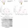
a, The SpyTag–SpyCatcher technology enables spontaneous isopeptide bond formation of the amino group of lysine side chains and the carboxyl group of aspartate side chains. This allows Spy-tagged proteins such as ZUD to be coupled to HRP for immunoblotting, Fc for immunofluorescence and biotin for streptavidin-based enrichment (Extended Data Fig. 6a). b, GFP–ZUD pulldown of ARH3-KO cells (four 500-cm2 dishes) transfected with GFP–ZUD and treated with 2 mM H2O2 (30 min). One sample was processed and the elution was split in two as described in the scheme (top). One half was treated with 1 M hydroxylamine (NH₂OH) and the other half was left untreated (−). The ZUD–HRP signal is abolished after treating the elution with NH₂OH for 2 h. This treatment preserves the mono-ADPr pattern (AbD43647–HRP-coupled) and the overall ubiquitin signal. Shown is a representative result from three independent experiments. c, Streptavidin pulldown of biotin-coupled ZUD from untreated, DNA-damage-treated (2 mM H2O2, 30 min) or DNA-damage-treated and olaparib-treated (1 µM) untransfected U2OS WT and untransfected ARH3-KO cells (two 15-cm dishes per condition were used). Immunoblots of the elutions for ZUD–HRP, AbD43647–HRP-coupled and PARP1 antibodies reveal a DNA-damage-dependent increase in ZUD–HRP signal and in mono-ADPr and PARP1. The bands likely to correspond to PARP1, PARP1 ADPr and PARP1 ADPrUb are labeled on each elution blot. An 8% Bis–Tris gel was used for the anti-PARP1 and AbD43647–HRP-coupled blots. A 4–12% Bis–Tris gradient gel was used for the ZUD–HRP-coupled blot. Shown is a representative result from three independent experiments. d, GFP–ZUD pulldown of ARH3-KO cells (eight 500-cm2 dishes) transfected with GFP–ZUD and treated with 2 mM H2O2 (30 min). The elution was split in two and half of it was treated with hydroxylamine (NH₂OH) as described in b. The H3 blot reveals NH2OH-sensitive ADPr-monoUb and ADPr-diUb bands. A 20% Bis–Tris gel was used to better resolve H3. A 4–12% Bis–Tris gradient gel was used for the ZUD–HRP-coupled blot. Following the EDTA elution, GFP–ZUD was eluted by heating the beads at 95 °C for 15 min. Shown is a representative result from four independent experiments. In bd, EDTA elution was performed for 5 min at room temperature. Source data
Extended Data Fig. 1
Extended Data Fig. 1. RNF114 interactors screen with GFP-RNF114 WT and C176A mutant inducible cell lines.
(a) GFP immunoblot with and without 24-hour doxycycline induction in inducible RNF114 KO U2OS cells complemented with GFP-RNF114 WT or GFP-RNF114 C176A expression. (b) Correlation analysis output of DIA-NN, showing the sample correlation within and between the conditions. A general separation of the samples belonging to different conditions is visible. (c) PCA analysis confirming the clear separation of samples from different conditions during the GFP-Pulldown. (d) Volcano plot showing the log2-fold change of the interactors identified in H2O2-treated against untreated GFP-RNF114 C176A overexpressing cells. (e) Comparison of interactors in H2O2-treated cells either overexpressing GFP-RNF114 WT or GFP-RNF114 C176A. (d,e) The interactors stated in Fig. 1 are marked if significant. Statistical analysis was performed using the limma’s two-sided moderated t-test. Adjusted P values were calculated using the Benjamini-Hochberg method to correct for multiple testing. The red dashed line in (d,e) displays significance with adjusted P values < 0.05 as -log10(adj. P value) >1.3. (f) Heatmaps showing the log2-fold change of the interactors stated in Fig. 1 (above) and the log2-fold change of selected Deubiquitinases (below) across all comparisons. Source data
Extended Data Fig. 2
Extended Data Fig. 2. Initial identification of ADP-ribosyl-ubiquitylation sites and EDTA elution conditions.
(a) HCD spectrum of ADP-ribosyl-ubiquitylation identified on HMGA1. (b) HCD spectrum of H2B showing the diagnostic ions of ADP-ribosyl-ubiquitylation. (a,b) Elution: 8 M Urea for 10 min at 37 °C. Four 10 cm dishes of U2OS WT cells transfected with GFP-ZUD were treated with 2 mM H2O2 (30 min). (c) Optimization of the EDTA elution temperature (4 °C, RT and 37 °C) and timing (30 seconds and 15 min). Shown is a representative result from three independent experiments. Source data
Extended Data Fig. 3
Extended Data Fig. 3. Additional spectra of peptides modified with ADP-ribosyl-ubiquitylation.
a,b) HCD and ETD spectra of ADP-ribosyl-ubiquitylation on serine 519 of PARP1. (c,d) HCD and ETD spectra of ADP-ribosyl-ubiquitylation on serine 6 of H2B. All shown HCD spectra contain the diagnostic ions of ADP-ribosyl-ubiquitylation. The ETD spectra do not contain diagnostic ions as the modification stays intact and is not fragmented, thus allowing the localization of the modification site. These spectra illustrate that ETD data is critical to localize the ADP-ribosyl-ubiquitylation on peptides. However, the presence of ADP-ribosyl-ubiquitylation can be validated by diagnostic ions using HCD. This illustrates how HCD and ETD data can complement each other. (a-d) EDTA elution: 15 min at 37 °C. Two 500-cm2 dishes of ARH3 KO cells transfected with GFP-ZUD were treated with 2 mM H2O2 (30 min).
Extended Data Fig. 4
Extended Data Fig. 4. Additional spectra of histone ADP-ribosyl-ubiquitylation marks.
(a,b) HCD and ETD spectra of ADP-ribosyl-ubiquitylation on serine 10 of H3. (c,d) HCD spectra of ADP-ribosyl-ubiquitylation likely on H3 serine 10 (c) or serine 28 (d) co-occurring with di-methylation of lysine 9 and lysine 36, respectively. (b) EDTA elution: 15 min at 37 °C. ARH3 KO cells (two 500-cm2 dishes) transfected with GFP-ZUD were treated with 2 mM H2O2 (30 min). (a,c,d) EDTA elution: 20 min at RT and ArgC digested. ARH3 KO cells (three 500-cm2 dishes) transfected with GFP-ZUD were treated with 2 mM H2O2 (30 min).
Extended Data Fig. 5
Extended Data Fig. 5. In vitro reactions using DTX2 and DTX3L to catalyze ADP-ribosyl-ubiquitylation on H3S10ADPr peptide or nucleosome.
(a) HCD spectra of the in vitro reaction with DTX2 RING-DTC domains and biotinylated ADP-ribosylated H3 peptide modified with ADP-ribosyl-ubiquitylation on serine 10. (b) Production of site-specific H3S10ADPr nucleosomes (adapted from and described in). (c) HCD and (d) ETD spectra of the in vitro reaction with DTX3L RING-DTC domains and biotinylated ADP-ribosylated H3 nucleosomes modified with ADP-ribosyl-ubquitylation on serine 10.
Extended Data Fig. 6
Extended Data Fig. 6. Coupling of SpyCatcher in different formats to the SpyTag-ZUD and specificity of ZUD.
(a) Scheme of the SpyCatcher-SpyTag reaction and the bivalent antibody-like reagent. (b) The coupling was analysed by Coomassie staining. On the left, the Biotin SpyCatcher coupling to the SpyTag-ZUD is shown. On the right, the Fc SpyCatcher coupling to the SpyTag-ZUD. Both gels display the uncoupled ZUD (SpyTag-ZUD) as input, the respective uncoupled SpyCatcher (Catcher), and the result of the coupling (coupled). 2 µg of each sample were loaded. (c) Signal of HRP-ZUD reagent in whole cell lysates of untreated, H2O2-treated and Olaparib + H2O2-treated ARH3 KO cells from a replicate of the experiment shown in Fig. 5c. Shown is a representative result from at least three independent experiments. (d) GFP-ZUD pulldown of ARH3 KO cells (four 500 cm2 dishes) transfected with GFP-ZUD and treated with 2 mM H2O2 (30 min). Half of the EDTA elution (5 min RT) was heated to 95 °C for 5 min. The ZUD-HRP signal is abolished after boiling. This treatment preserves the mono-ADPr pattern (AbD43647 HRP-coupled) and the overall ubiquitin signal. Shown is a representative result from three independent experiments. Source data
Extended Data Fig. 7
Extended Data Fig. 7. Specificity of ZUD and pulldown of histone H2B.
(a) GFP pulldown of ARH3 KO cells (two 500 cm2 dishes per condition) transfected with GFP-ZUD or GFP-zfDi19 and treated with 2 mM H2O2 (30 min). The elutions (5 min RT) of both pulldowns were immunoblotted for ZUD HRP-coupled, AbD43647 HRP-coupled and ubiquitin antibody. zfDi19 (lacking UIM) shows negligible signal for AbD43647 HRP-coupled, ubiquitin and HRP-ZUD compared to ZUD (zfDi19-UIM), showing the specificity of ZUD for ADP-ribosyl-ubiquitylation. Shown is a representative result from three independent experiments. The GFP input and 95 °C after EDTA elution blots were cropped for clarity. The asterisk (*) indicates the position where the image was cut. Full-length blots are provided in the Source Data of this figure. (b) GFP-ZUD pulldown of ARH3 KO cells (eight 500 cm2 dishes) transfected with GFP-ZUD and treated with 2 mM H2O2 (30 min). Immunoblotting the elution with an H2B antibody reveals an NH₂OH-sensitive ADPrUb band. 20% Bis-Tris gel was used to better resolve H2B. Shown is a representative result from three independent experiments. (a,b) Following the EDTA elution, GFP-ZUD was eluted by heating the beads at 95 °C for 15 min. Source data

References

    1. Walsh, C. T., Garneau-Tsodikova, S. & Gatto, G. J. Jr. Protein posttranslational modifications: the chemistry of proteome diversifications. Angew. Chem. Int. Ed. Engl.44, 7342–7372 (2005). - PubMed
    1. Dikic, I. & Schulman, B. A. An expanded lexicon for the ubiquitin code. Nat. Rev. Mol. Cell Biol.24, 273–287 (2023). - PMC - PubMed
    1. Suskiewicz, M. J., Prokhorova, E., Rack, J. G. M. & Ahel, I. ADP-ribosylation from molecular mechanisms to therapeutic implications. Cell186, 4475–4495 (2023). - PMC - PubMed
    1. Gupte, R., Liu, Z. & Kraus, W. L. PARPs and ADP-ribosylation: recent advances linking molecular functions to biological outcomes. Genes Dev.31, 101–126 (2017). - PMC - PubMed
    1. Luscher, B. et al. ADP-ribosyltransferases, an update on function and nomenclature. FEBS J.289, 7399–7410 (2022). - PMC - PubMed

MeSH terms

LinkOut - more resources