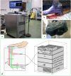
In vivo photoacoustic tomography of porcine abdominal organs using Fabry-Pérot sensing integrated platform
- PMID: 40634645
- PMCID: PMC12241532
- DOI: 10.1186/s41747-025-00601-1
In vivo photoacoustic tomography of porcine abdominal organs using Fabry-Pérot sensing integrated platform
Abstract
Objective: To evaluate in vivo a fully integrated photoacoustic tomography imaging system based on Fabry-Pérot ultrasound sensing method applied on porcine abdominal organs. This approach could be used by surgeons during intraoperative clinical procedures.
Methods: The photoacoustic imaging system was fully integrated into a single structure, and the detection technology was based on a Fabry-Pérot interferometer. The detection probe connected to the imaging system was applied directly to the organs of a male "large white" Sus scrofa pig weighing 80 kg, either manually or using a stand, with or without a gel interface. All experiments were performed in compliance with EU Directive 2010/63/EU on animal experimentation (APAFiS #31507).
Results: All intraperitoneal and retroperitoneal organs were evaluated using photoacoustic imaging. The evaluation of both hollow and solid organs was successfully conducted with consistent three-dimensional image quality. We demonstrate the system's ability to image blood vessels with diameters ranging from several millimeters down to less than 100 µm. Macroscopic evaluation of the organs using photoacoustic tomography imaging did not reveal any damage or burns caused by the excitation laser.
Conclusion: To our knowledge, this is the first reported imaging session of abdominal organs in an in vivo porcine model, performed using a photoacoustic tomography system with Fabry-Pérot interferometer detection. We present a high-resolution photoacoustic tomography system that is closer to routine clinical translation, thanks to a fully integrated system.
Relevance statement: Photoacoustic evaluation of organs using a fully integrated system could become a valuable tool for surgical teams for intraprocedural assessment of vascularization.
Key points: Photoacoustic imaging visualizes blood vessels without contrast agents or ionizing radiation. Photoacoustic imaging systems detect blood vessels ranging from millimeters to 100 µm. Fully integrated photoacoustic imaging systems are autonomously operable by surgical teams.
Keywords: Anatomy; Animal; Disease model; Equipment; Photoacoustic techniques.
© 2025. The Author(s).
Conflict of interest statement
Declarations. Ethics approval and consent to participate: The French Ministry of Research approved the research protocol (APAFiS nos 31507). All experiments were conducted in accordance with the ARRIVE 2.0 recommendations and the European Directive 2010/63/EU on animal experimentation. Competing interests: F Richard, A Vrignaud, D Gasteau, and A Biallais were, respectively, the chief executive officer, chief technical officer, and employees of DeepColor SAS. DeepColor SAS is a French limited company commercializing photoacoustic tomography systems. The other authors have no conflicts of interest to disclose as described by European Radiology Experimental. Consent for publication: All experiments were conducted in accordance with the ARRIVE 2.0 recommendations and the European Directive 2010/63/EU on animal experimentation.
Figures


Similar articles
-
Comparison of Two Modern Survival Prediction Tools, SORG-MLA and METSSS, in Patients With Symptomatic Long-bone Metastases Who Underwent Local Treatment With Surgery Followed by Radiotherapy and With Radiotherapy Alone.Clin Orthop Relat Res. 2024 Dec 1;482(12):2193-2208. doi: 10.1097/CORR.0000000000003185. Epub 2024 Jul 23. Clin Orthop Relat Res. 2024. PMID: 39051924
-
Contrast-enhanced ultrasound using SonoVue® (sulphur hexafluoride microbubbles) compared with contrast-enhanced computed tomography and contrast-enhanced magnetic resonance imaging for the characterisation of focal liver lesions and detection of liver metastases: a systematic review and cost-effectiveness analysis.Health Technol Assess. 2013 Apr;17(16):1-243. doi: 10.3310/hta17160. Health Technol Assess. 2013. PMID: 23611316 Free PMC article.
-
Magnetic resonance perfusion for differentiating low-grade from high-grade gliomas at first presentation.Cochrane Database Syst Rev. 2018 Jan 22;1(1):CD011551. doi: 10.1002/14651858.CD011551.pub2. Cochrane Database Syst Rev. 2018. PMID: 29357120 Free PMC article.
-
Signs and symptoms to determine if a patient presenting in primary care or hospital outpatient settings has COVID-19.Cochrane Database Syst Rev. 2022 May 20;5(5):CD013665. doi: 10.1002/14651858.CD013665.pub3. Cochrane Database Syst Rev. 2022. PMID: 35593186 Free PMC article.
-
Systemic pharmacological treatments for chronic plaque psoriasis: a network meta-analysis.Cochrane Database Syst Rev. 2021 Apr 19;4(4):CD011535. doi: 10.1002/14651858.CD011535.pub4. Cochrane Database Syst Rev. 2021. Update in: Cochrane Database Syst Rev. 2022 May 23;5:CD011535. doi: 10.1002/14651858.CD011535.pub5. PMID: 33871055 Free PMC article. Updated.
References
-
- Mesnard B, Branchereau J, Prudhomme T (2025) Photoacoustic tomography assessments during ex vivo normothermic perfusion. A novel and noninvasive modality to evaluate endothelial integrity. Transplantation. 10.1097/TP.0000000000005342 - PubMed
MeSH terms
LinkOut - more resources
Full Text Sources
