Direct observation of long-lived radical pair between flavin and guanine in single- and double-stranded DNA-oligomers
- PMID: 40634712
- PMCID: PMC12241510
- DOI: 10.1038/s42004-025-01596-x
Direct observation of long-lived radical pair between flavin and guanine in single- and double-stranded DNA-oligomers
Abstract
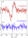
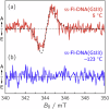
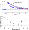
The mechanism by which cryptochrome (CRY) proteins are capable of sensing weak magnetic fields (e.g., the geomagnetic field: ~50 μT) was suggested to be mediated by spin-correlated radical pairs (SCRPs) comprising a flavin adenine dinucleotide (FAD) radical and a tryptophan (Trp) radical which are formed simultaneously by light-induced electron transfer (ET). Here, we provide evidence for direct photoinduced ET that leads to long-lived SCRPs comprising a flavin (Fl) radical and a guanine (G) radical in flavin-tethered single- and double-stranded DNA oligomers by using time-resolved electron paramagnetic resonance (TREPR) spectroscopy. Transient absorption (TA) spectroscopy and its magnetic field effect (MFE) identified RP generation via a triplet-state precursor, in contrast to RP generation via a singlet-state precursor in CRY. Our findings of RPs in Fl-DNA oligomers having microsecond-long lifetimes and capable of exerting a large MFE at room temperature may significantly impact on our understanding of biological magnetoreception.
© 2025. The Author(s).
Conflict of interest statement
Competing interests: The authors declare no competing interests.
Figures


Similar articles
-
Electron hopping in cryptochrome: Implications for radical pair magnetoreception and the role of the fourth tryptophan.J Chem Phys. 2025 Jul 14;163(2):024110. doi: 10.1063/5.0278806. J Chem Phys. 2025. PMID: 40631818
-
Decrypting the Nonadiabatic Photoinduced Electron Transfer Mechanism in Light-Sensing Cryptochrome.ACS Cent Sci. 2025 May 30;11(7):1071-1082. doi: 10.1021/acscentsci.5c00376. eCollection 2025 Jul 23. ACS Cent Sci. 2025. PMID: 40726782 Free PMC article.
-
Systemic pharmacological treatments for chronic plaque psoriasis: a network meta-analysis.Cochrane Database Syst Rev. 2017 Dec 22;12(12):CD011535. doi: 10.1002/14651858.CD011535.pub2. Cochrane Database Syst Rev. 2017. Update in: Cochrane Database Syst Rev. 2020 Jan 9;1:CD011535. doi: 10.1002/14651858.CD011535.pub3. PMID: 29271481 Free PMC article. Updated.
-
The effect of sample site and collection procedure on identification of SARS-CoV-2 infection.Cochrane Database Syst Rev. 2024 Dec 16;12(12):CD014780. doi: 10.1002/14651858.CD014780. Cochrane Database Syst Rev. 2024. PMID: 39679851 Free PMC article.
-
Systemic pharmacological treatments for chronic plaque psoriasis: a network meta-analysis.Cochrane Database Syst Rev. 2021 Apr 19;4(4):CD011535. doi: 10.1002/14651858.CD011535.pub4. Cochrane Database Syst Rev. 2021. Update in: Cochrane Database Syst Rev. 2022 May 23;5:CD011535. doi: 10.1002/14651858.CD011535.pub5. PMID: 33871055 Free PMC article. Updated.
References
-
- Mohseni, M., Omar, Y., Engel, G. S. & Plenio, M. B. and references therein. Quantum Effects in Biology. (Cambridge Univ. Press, Cambridge, 2014).
-
- Blaustein, G. S., Lewis, F. D. & Burin, A. L. Kinetics of charge separation in poly(A)-poly(T) DNA hairpins. J. Phys. Chem. B114, 6732–6739 (2010). - PubMed
-
- Giese, B., Amaudrut, J., Köhler, A.-K., Spormann, M. & Wessely, S. Direct observation of hole transfer through DNA by hopping between adenine bases and by tunnelling. Nature412, 318–320 (2001). - PubMed
Grants and funding
LinkOut - more resources
Full Text Sources

